. 2012 Feb 3;287(6):4121-8.
doi: 10.1074/jbc.M111.285395. Epub 2011 Dec 6.

Structural and functional insights into the DNA replication factor Cdc45 reveal an evolutionary relationship to the DHH family of phosphoesterases

Affiliations

Structural and functional insights into the DNA replication factor Cdc45 reveal an evolutionary relationship to the DHH family of phosphoesterases

Ivet Krastanova et al. J Biol Chem. .

Abstract

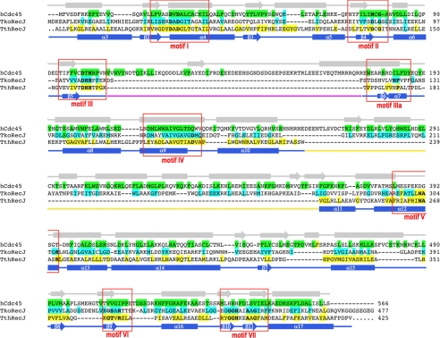
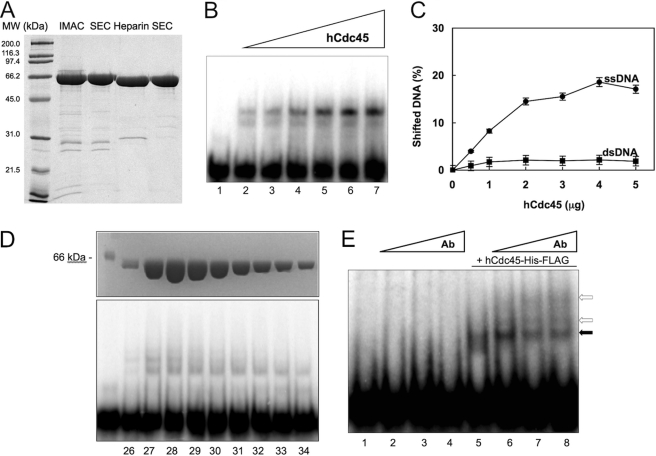
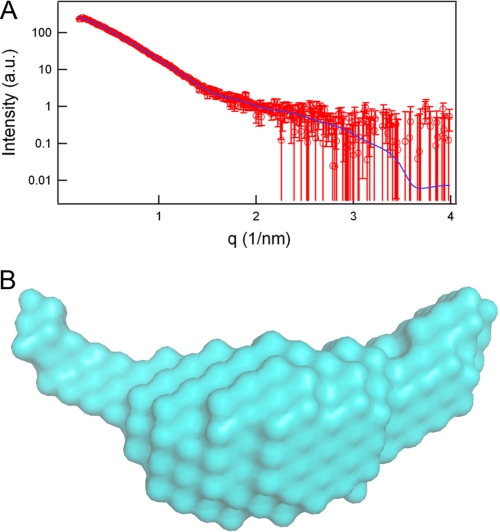
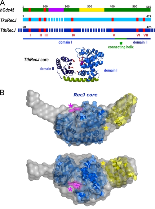
Cdc45 is an essential protein conserved in all eukaryotes and is involved both in the initiation of DNA replication and the progression of the replication fork. With GINS, Cdc45 is an essential cofactor of the Mcm2-7 replicative helicase complex. Despite its importance, no detailed information is available on either the structure or the biochemistry of the protein. Intriguingly, whereas homologues of both GINS and Mcm proteins have been described in Archaea, no counterpart for Cdc45 is known. Herein we report a bioinformatic analysis that shows a weak but significant relationship among eukaryotic Cdc45 proteins and a large family of phosphoesterases that has been described as the DHH family, including inorganic pyrophosphatases and RecJ ssDNA exonucleases. These enzymes catalyze the hydrolysis of phosphodiester bonds via a mechanism involving two Mn(2+) ions. Only a subset of the amino acids that coordinates Mn(2+) is conserved in Cdc45. We report biochemical and structural data on the recombinant human Cdc45 protein, consistent with the proposed DHH family affiliation. Like the RecJ exonucleases, the human Cdc45 protein is able to bind single-stranded, but not double-stranded DNA. Small angle x-ray scattering data are consistent with a model compatible with the crystallographic structure of the RecJ/DHH family members.


Figures

FIGURE 1.
FIGURE 1.
Sequence alignment between hCdc45, RecJCdc45 from T. kodakaraensis, and RecJ from T. thermophilus. The alignment presented here is based on an extended multiple alignment using 15 eukaryotic Cdc45 sequences, 16 archaeal sequences, and 10 bacterial RecJ sequences, selected from evolutionary diverse organisms. Only the RecJ core (residues 50–425, comprising domains I and II) has been used in the alignment. Residues that are conserved in more than 70% of the eukaryotic, archaeal, and bacterial sequences are highlighted in green, cyan, and yellow, respectively. The following groups of amino acid residues were considered similar: Asp/Glu, Lys/Arg, Phe/Tyr, Ser/Thr, Gly/Ala, and Val/Leu/Ile/Met. The position of the secondary structural elements in the T. thermophilus RecJ crystal structure (PDB code: 2ZXP) is indicated at the bottom, whereas the predicted secondary structure for human Cdc45 is shown at the top. Secondary structure elements are named according to the TthRecJ nomenclature (28). The position of the characteristic RecJ motifs is shown by red boxes, with the residues conserved highlighted in bold. The alignment was carried out using the multiple sequence alignment program MUSCLE (41) and manually modified to take into account the structural constraints, and the results of the -fold recognition/threading algorithms. Up to motif IV the similarity is strong enough to be detected based on sequence alone, whereas the second half of the alignment relies on the threading data, which identify similarity patterns in the absence of high sequence homology, as exemplified by the conservation of the patterns of hydrophobic residues and the excellent match between RecJ secondary structure elements and the prediction for Cdc45. An insertion unique to eukaryotic Cdc45 orthologues and containing many charged amino acid residues is shown in magenta. The putative helical insertion present in both archaeal and eukaryotic proteins is shown in yellow.
FIGURE 2.
FIGURE 2.
Biochemical characterization of recombinant human Cdc45. A, purification of hCdc45. SDS-PAGE analysis of samples throughout the purification protocol (see detailed description under “Experimental Procedures”), starting from the protein obtained after the first step of Ni-affinity purification (IMAC (immobilized metal-ion affinity chromatography)), followed by size-exclusion chromatography (SEC); the protein after cleavage of the His6-FLAG tag using TEV protease and purification over a heparin column (Heparin) to a final round of SEC. B, DNA-binding activity of hCdc45. Example of an EMSA on single-stranded DNA is shown. The assay was carried out with increasing concentrations of hCdc45 (0.5, 1, 2, 3, 4, and 5 μg of protein were present in the mixtures loaded into the lanes from 2 to 7). A radiolabeled 56-mer DNA was used as a ligand (see the text for details). A control mixture without protein was run on lane 1. C, single-stranded versus double-stranded DNA binding. Shifted DNA (either in single- (●) or double-stranded (■) form) is reported versus the amount of hCdc45. Experiments were performed in triplicate, and the results are averaged. Curves represent best fits to the data points. The error bars on the graphs are the ±S.E. D, gel-filtration analysis of hCdc45 and EMSAs of the corresponding peak fractions. Gel-filtration chromatography of purified hCdc45 was performed using a Bio-Sil SEC-250 column (Bio-Rad) as described under “Experimental Procedures.” Peak fractions were analyzed by SDS-PAGE (5 μl/fraction) and used in a gel shift experiment (1 μl/fraction). E, EMSA with hCdc45-His-FLAG in the presence of anti-FLAG antibody. The assays were carried out by adding increasing amounts of a monoclonal anti-FLAG antibody (0.5, 1, and 2 μg, lanes 2 and 6, 3 and 7, and 4 and 8, respectively) into mixtures containing the single-stranded DNA probe with hCdc45-His-FLAG (lanes 6–8; 5 μg of protein) or without the recombinant protein (lanes 2–4; see text for details). A black arrow indicates the Cdc45-DNA complex, whereas the white arrows identify the ternary complexes with the anti-FLAG antibody.
FIGURE 3.
FIGURE 3.
Small angle x-ray scattering data. A, the experimental SAXS profile (log intensity as a function of the momentum transfer) of hCdc45 (red points) is compared with the theoretical scattering curves calculated from the ab initio model (blue line). B, final model reconstructed from the scattering curve.
FIGURE 4.
FIGURE 4.
SAXS data are consistent with a RecJ-like fold. A, a schematic diagram summarizing the result of the bioinformatic analysis and showing the relationship between eukaryotic Cdc45 proteins, archaeal RecJCdc45, and bacterial RecJ single-stranded DNA exonucleases. The RecJ motifs are shown in red, the Cdc45 charged insertion is in magenta, and the helical archaeal/eukaryotic insertion is in yellow. The crystal structure of the core of T. thermophilus RecJ (PDB code: 2ZXP, domains I and II) is shown below the diagram, with domain I in blue, domain II in dark blue, and the connecting helix in green. The conserved residues in the seven RecJ motifs are shown in red. B, the ab initio calculated SAXS model for hCdc45 (depicted as gray light spheres) is superimposed to the crystal structure of the core of T. thermophilus RecJ, in blue. Highlighted in magenta and indicated by a magenta asterisk is the putative position of the insertion, which is unique to the eukaryotic Cdc45 orthologues; highlighted in yellow and indicated by a yellow asterisk is the position of the helical bundle insertion that is common to both archaeal and eukaryotic proteins (see Fig. 1). As an example, the helical domain of the acyl-CoA-binding protein (PDB code: 2FDQ) has been fitted to the map, consistently with the results of the threading algorithms. The two views are roughly related by a 90° rotation around a horizontal axis.

Similar articles

Cited by

References

    1. Aparicio O. M., Weinstein D. M., Bell S. P. (1997) Components and dynamics of DNA replication complexes in S. cerevisiae. Redistribution of MCM proteins and Cdc45p during S phase. Cell 91, 59–69 - PubMed
    1. Owens J. C., Detweiler C. S., Li J. J. (1997) CDC45 is required in conjunction with CDC7/DBF4 to trigger the initiation of DNA replication. Proc. Natl. Acad. Sci. U.S.A. 94, 12521–12526 - PMC - PubMed
    1. Uchiyama M., Arai K., Masai H. (2001) Sna41goa1, a novel mutation causing G1/S arrest in fission yeast, is defective in a CDC45 homolog and interacts genetically with polα. Mol. Genet. Genomics 265, 1039–1049 - PubMed
    1. Mimura S., Takisawa H. (1998) Xenopus Cdc45-dependent loading of DNA polymerase α onto chromatin under the control of S-phase Cdk. EMBO J. 17, 5699–5707 - PMC - PubMed
    1. Pacek M., Walter J. C. (2004) A requirement for MCM7 and Cdc45 in chromosome unwinding during eukaryotic DNA replication. EMBO J. 23, 3667–3676 - PMC - PubMed

Publication types

MeSH terms

LinkOut - more resources