Corticostriatal-limbic gray matter morphology in adolescents with self-reported exposure to childhood maltreatment
- PMID: 22147775
- PMCID: PMC3607102
- DOI: 10.1001/archpediatrics.2011.565
Corticostriatal-limbic gray matter morphology in adolescents with self-reported exposure to childhood maltreatment
Abstract
Objective: To study the relationship between self-reported exposure to childhood maltreatment (CM) and cerebral gray matter (GM) morphology in adolescents without psychiatric diagnoses.
Design: Associations were examined between regional GM morphology and exposure to CM (measured using a childhood trauma self-report questionnaire for physical, emotional, and sexual abuse and for physical and emotional neglect).
Setting: University hospital.
Participants: Forty-two adolescents without psychiatric diagnoses.
Main outcome measures: Correlations between childhood trauma self-report questionnaire scores and regional GM volume were assessed in voxel-based analyses of structural magnetic resonance images. Relationships among GM volume, subtypes of exposure to CM, and sex were explored.
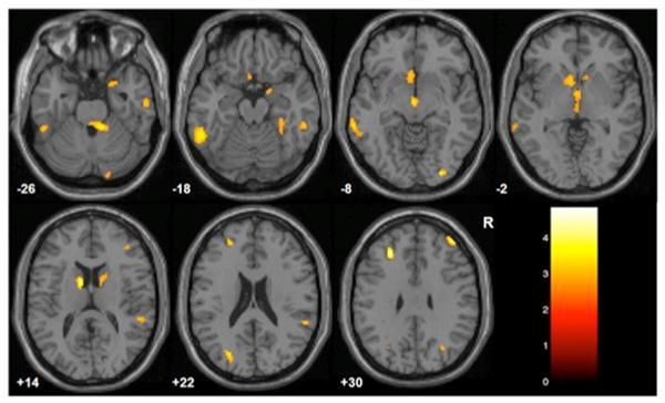
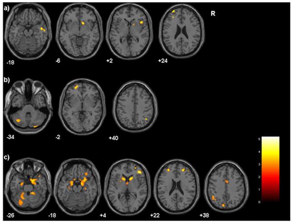
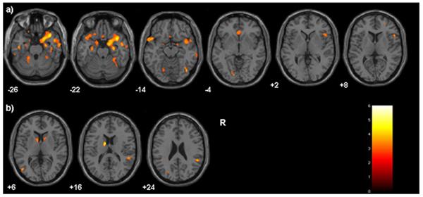
Results: Childhood trauma self-report questionnaire total scores correlated negatively (P < .005) with GM volume in prefrontal cortex, striatum, amygdala, sensory association cortices, and cerebellum. Physical abuse, physical neglect, and emotional neglect were associated with rostral prefrontal reductions. Decreases in dorsolateral and orbitofrontal cortices, insula, and ventral striatum were associated with physical abuse. Decreases in cerebellum were associated with physical neglect. Decreases in dorsolateral, orbitofrontal, and subgenual prefrontal cortices, striatum, amygdala, hippocampus, and cerebellum were associated with emotional neglect. Decreases in the latter emotion regulation regions were also associated with childhood trauma self-report questionnaire scores in girls, while caudate reductions (which may relate to impulse dyscontrol) were seen in boys.
Conclusions: Exposure to CM was associated with corticostriatal-limbic GM reductions in adolescents. Even if adolescents reporting exposure to CM do not present with symptoms that meet full criteria for psychiatric disorders, they may have corticostriatal-limbic GM morphologic alterations that place them at risk for behavioral difficulties. Vulnerabilities may be moderated by sex and by subtypes of exposure to CM.
Figures
Comment in
-
Conceptual and methodological issues in neuroimaging studies of the effects of child maltreatment.Arch Pediatr Adolesc Med. 2011 Dec;165(12):1133-4. doi: 10.1001/archpediatrics.2011.1046. Arch Pediatr Adolesc Med. 2011. PMID: 22147781 Free PMC article. No abstract available.
Similar articles
-
Neuroanatomical changes associated with conduct disorder in boys: influence of childhood maltreatment.Eur Child Adolesc Psychiatry. 2022 Apr;31(4):601-613. doi: 10.1007/s00787-020-01697-z. Epub 2021 Jan 4. Eur Child Adolesc Psychiatry. 2022. PMID: 33398650
-
Childhood maltreatment and identity diffusion among inpatient adolescents: The role of reflective function.J Adolesc. 2019 Oct;76:65-74. doi: 10.1016/j.adolescence.2019.08.002. Epub 2019 Aug 28. J Adolesc. 2019. PMID: 31472427
-
Sex differences of gray matter morphology in cortico-limbic-striatal neural system in major depressive disorder.J Psychiatr Res. 2013 Jun;47(6):733-9. doi: 10.1016/j.jpsychires.2013.02.003. Epub 2013 Mar 1. J Psychiatr Res. 2013. PMID: 23453566 Free PMC article.
-
Annual Research Review: Enduring neurobiological effects of childhood abuse and neglect.J Child Psychol Psychiatry. 2016 Mar;57(3):241-66. doi: 10.1111/jcpp.12507. Epub 2016 Feb 1. J Child Psychol Psychiatry. 2016. PMID: 26831814 Free PMC article. Review.
-
Understanding heterogeneity in grey matter research of adults with childhood maltreatment-A meta-analysis and review.Neurosci Biobehav Rev. 2016 Oct;69:299-312. doi: 10.1016/j.neubiorev.2016.08.011. Epub 2016 Aug 13. Neurosci Biobehav Rev. 2016. PMID: 27531235 Review.
Cited by
-
Corporal Punishment and Elevated Neural Response to Threat in Children.Child Dev. 2021 May;92(3):821-832. doi: 10.1111/cdev.13565. Epub 2021 Apr 9. Child Dev. 2021. PMID: 33835477 Free PMC article.
-
Left amygdala structure mediates longitudinal associations between exposure to threat and long-term psychiatric symptomatology in youth.Hum Brain Mapp. 2022 Sep;43(13):4091-4102. doi: 10.1002/hbm.25904. Epub 2022 May 18. Hum Brain Mapp. 2022. PMID: 35583310 Free PMC article.
-
Hippocampal and parahippocampal volumes vary by sex and traumatic life events in children.J Psychiatry Neurosci. 2020 Jul 1;45(4):288-297. doi: 10.1503/jpn.190013. J Psychiatry Neurosci. 2020. PMID: 32078279 Free PMC article.
-
Early Adverse Experiences and the Developing Brain.Neuropsychopharmacology. 2016 Jan;41(1):177-96. doi: 10.1038/npp.2015.252. Epub 2015 Sep 3. Neuropsychopharmacology. 2016. PMID: 26334107 Free PMC article. Review.
-
The sexually dimorphic impact of maltreatment on cortical thickness, surface area and gyrification.J Neural Transm (Vienna). 2016 Sep;123(9):1069-83. doi: 10.1007/s00702-016-1523-8. Epub 2016 Feb 27. J Neural Transm (Vienna). 2016. PMID: 26922372 Free PMC article.
References
-
- U.S. Department of Health and Human Services, Administration for Children and Families, Children's Bureau Child Maltreatment: Reports from the States to the National Child Abuse and Neglect Data System. 2010 http://www.acf.hhs.gov/programs/cb/stats_research/index.htm#can.
-
- Kaffman A, Meaney MJ. Neurodevelopmental sequelae of postnatal maternal care in rodents: clinical and research implications of molecular insights. J Child Psychol Psychiatry. 2007;48:224–244. - PubMed
-
- Monroy E, Hernandez-Torres E, Flores G. Maternal separation disrupts dendritic morphology of neurons in prefronal cortex, hippocampus, and nucleus accumbens in male rat offspring. J Chem Neuroanat. 2010 In Press. - PubMed
-
- Bremner JD, Vermetten E. Stress and development. behavioral and biological consequences. Dev Psychopathol. 2001;13:473–489. - PubMed
-
- Bremner JD, Vythilingam M, Vermetten E, et al. MRI and PET study of deficits in hippocampal structure and function in women with childhood sexual abuse and posttraumatic stress disorder. Am J Psychiatry. 2003;160:924–932. - PubMed
Publication types
MeSH terms
Grants and funding
- R01MH070902/MH/NIMH NIH HHS/United States
- K01 MH086621/MH/NIMH NIH HHS/United States
- RL5DA024858/DA/NIDA NIH HHS/United States
- R01MH69747/MH/NIMH NIH HHS/United States
- UL1 DE019586/DE/NIDCR NIH HHS/United States
- K05DA020091/DA/NIDA NIH HHS/United States
- RL1 DA024856/DA/NIDA NIH HHS/United States
- RL1DA024856/DA/NIDA NIH HHS/United States
- R01 MH069747/MH/NIMH NIH HHS/United States
- K01MH086621/MH/NIMH NIH HHS/United States
- R01 MH070902/MH/NIMH NIH HHS/United States
- UL1-DE19586/DE/NIDCR NIH HHS/United States
- K05 DA020091/DA/NIDA NIH HHS/United States
- PL1 DA024859/DA/NIDA NIH HHS/United States
- PL1-DA24859/DA/NIDA NIH HHS/United States
- RL5 DA024858/DA/NIDA NIH HHS/United States
LinkOut - more resources
Full Text Sources
Medical