. 2012 Feb 1;205(3):412-21.
doi: 10.1093/infdis/jir756. Epub 2011 Dec 5.

Immunization of mice with vibrio cholerae outer-membrane vesicles protects against hyperinfectious challenge and blocks transmission

Affiliations

Immunization of mice with vibrio cholerae outer-membrane vesicles protects against hyperinfectious challenge and blocks transmission

Anne L Bishop et al. J Infect Dis. .

Abstract

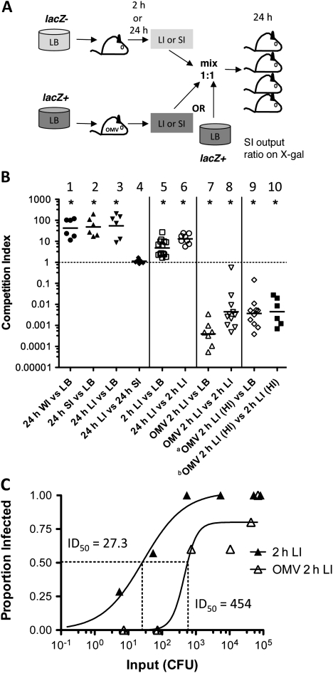
Background: Vibrio cholerae excreted by cholera patients is "hyperinfectious" (HI), which can be modeled by passage through infant mice. Immunization of adult female mice with V. cholerae outer-membrane vesicles (OMVs) passively protects suckling mice from challenge. Although V. cholerae is unable to colonize protected pups, the bacteria survive passage and have the potential to be transmitted to susceptible individuals. Here, we investigated the impact of OMV immunization and the HI state on V. cholerae transmission.

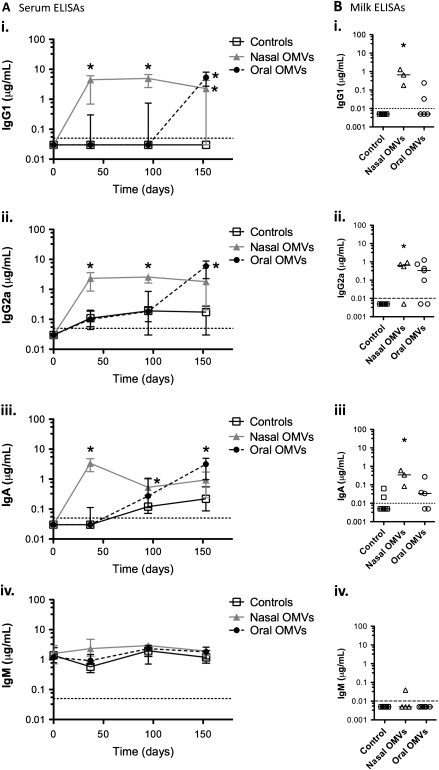
Methods: Neonatal mice suckled by OMV- or sham-immunized dams were challenged with HI V. cholerae. The infectivity of spatially and temporally separate V. cholerae populations obtained from infected naive or protected pups was tested. Recombination-based in vivo expression technology was used to assess virulence gene expression within these populations.

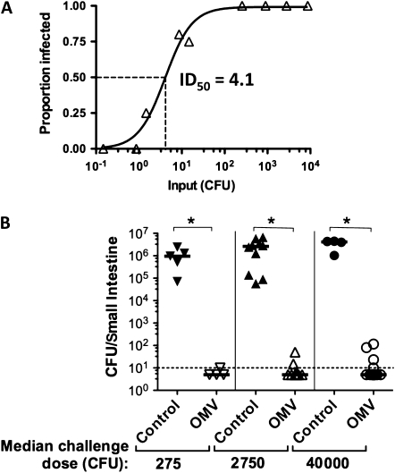
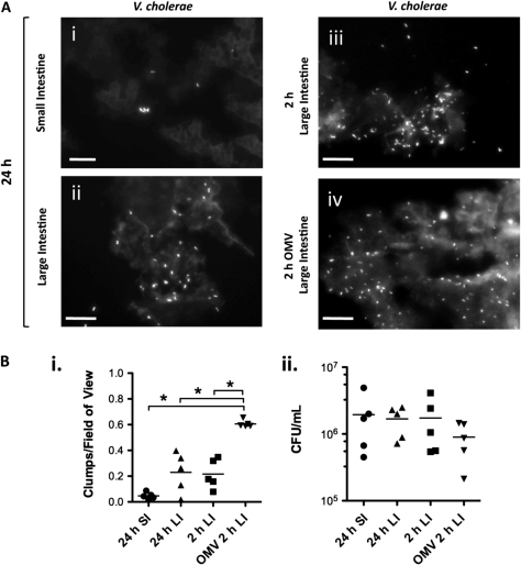
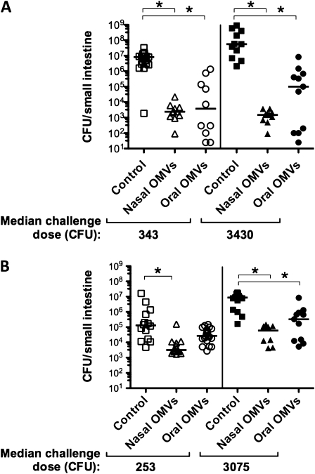
Results: OMV immunization significantly reduced colonization of neonates challenged with HI V. cholerae. Vibrio cholerae that had colonized the naive host was HI, whereas V. cholerae excreted by neonates born to OMV-immunized dams, although viable, was hypoinfectious and failed to fully induce virulence gene expression.

Conclusions: OMV immunization can significantly reduce the V. cholerae burden upon challenge with HI V. cholerae and can also block transmission from immune mice by reducing the infectivity of shed bacteria.


Figures

Figure 1.
Figure 1.
Challenge of outer-membrane vesicle (OMV)–immunized mice with hyperinfectious, mouse-passaged Vibrio cholerae. A, ID50 (the dose at which half the animals are infected) for secondary infection using, as inoculae, mouse-passaged V. cholerae E7946 recovered from primary infection small intestinal (SI) homogenates. Infected BALB/c neonates were returned to their dams postinoculation. Primary infection was with 105 E7946 from Luria-Bertani agar plates. Secondary infection was with SI homogenates diluted in saline to between 0.1 and 3 × 10⁴ colony-forming units (CFU)/50 μL challenge dose. Secondary infections were done in groups of 4–10 mice per challenge dose. Both primary and secondary infections were for 24 hours. The proportion of mice infected, with a colonization cutoff of >100 CFU, versus challenge dose is shown, from which the ID50 was calculated to be 4.1 (95% confidence interval, 2.200–5.995; R2 = 0.9814). B, Challenge of neonates born to sham- (control) or OMV-immunized dams with mouse-passaged V. cholerae was carried out with 3 challenge doses (median challenge doses indicated below the graph). Each symbol represents 1 neonate. Neonates from 3 mice are represented in each group. Bars = medians; dotted line = limit of detection. *P < .05 comparing pups born to controls and immunized mice infected with the same challenge dose (Mann–Whitney U tests). The anti-OMV enzyme-linked immunosorbent assay immunoglobulin G1 titers for immunized dams whose pups were used for this experiment ranged from 10.5 to 159 (median, 143) μg/mL.
Figure 2.
Figure 2.
Clumping of Vibrio cholerae within infected intestinal homogenates. Clumping of V. cholerae within infected mouse intestinal homogenates was assessed by visualization of formalin-fixed samples stained with fluorescein isothiocyanate–mouse anti-O1 V. cholerae antibody. A, Representative images of clumps within homogenates from small intestine (SI) (i) or large intestine (LI) (iiiv) harvested 24 hours (i, ii) or 2 hours (iii, iv) postinoculation and stained for V. cholerae. Scale bars = 20 μm. B, (i) Clumps of V. cholerae were scored in at least 20 fields of view with a ×60 objective from 2 samples taken from 5 fixed SI or LI homogenate samples that were harvested 2 hours or 24 hours postinoculation, as indicated. The number of clumps per field of view is shown. *P < .05 (1-way analysis of variance and post hoc t tests with Bonferroni adjustment). (ii) Viable counts (colony-forming units [CFU]/mL) for each homogenate, prepared as if for mouse infection, determined by serial dilution and plating prior to formaldehyde fixing. Each symbol represents 1 homogenate; bars = means. Abbreviation: OMV, outer membrane vesicle.
Figure 3.
Figure 3.
Immune responses of outer-membrane vesicle (OMV)–immunized Swiss Webster mice. Adult female Swiss Webster mice were immunized with three 25-μg doses of a 1:1 mixture of O1 Ogawa and O1 Inaba OMVs either intranasally (nasal, 10 mice) or by oral gavage (oral, 9 mice) at days 0, 14, and 28. Controls (19 mice) were cohoused with the immunized mice. Enzyme-linked immunosorbent assays (ELISAs) for serum and milk samples from these mice were carried out against O1 Ogawa OMVs. A, Serum levels of anti-O1 Ogawa OMV immunoglobulin (Ig) G1 μg/mL (i), IgG2a μg/mL (ii), IgA μg/mL (iii), and IgM μg/mL (iv) assessed by ELISA for preimmune serum (day 0) and serum samples taken at various days postimmunization, as indicated on the x-axis of each graph: day 37, before mating; day 95, after initial neonatal rice-water-stool challenges; day 153, after second round of challenge experiments. Sera from at least 6 mice per group were analyzed at each time point, except for day 153 when only 4 mice remained in the orally immunized group. Symbols = medians; error bars = interquartile range. *P < .05 compared with controls at the same time point (Kruskal–Wallis and Dunn tests). B, Levels of anti-O1 Ogawa OMV IgG1 μg/mL (i), IgG2a μg/mL (ii), IgA μg/mL (iii), and IgM μg/mL (iv) in milk extracted from the stomachs of pups born to OMV-immunized (nasally or orally delivered as indicated) or control mice. Each symbol represents milk pooled from the stomachs of at least 3 pups suckled by 1 dam; bars = medians. *P < .05 compared with controls (Kruskal–Wallis and Dunn tests).
Figure 4.
Figure 4.
Challenge of outer-membrane vesicle (OMV)–immunized Swiss Webster mice with rice-water-stool Vibrio cholerae. Neonates born to intranasally or orally O1 OMV-immunized or cohoused (control) dams were challenged with cholera patient rice-water-stool samples diluted to give the median challenge doses indicated. Challenges were carried out either (A) 62–76 days or (B) 143–149 days postimmunization of the dam. Each symbol represents viable counts in the small intestine of 1 challenged neonate; bars = medians. *P < .05 significantly lower viable counts compared with controls with the same challenge dose (Kruskal–Wallis and Dunn tests). Abbreviation: CFU, colony-forming units.
Figure 5.
Figure 5.
Infectivity of neonatal mouse host-passaged Vibrio cholerae 2 hours or 24 hours postinoculation and the impact of outer-membrane vesicle (OMV) immunization. A, Scheme for competitions between V. cholerae passaged through immunized (OMV) or unimmunized mice for 2 hours or 24 hours, or V. cholerae E7946 grown on Luria-Bertani (LB) plates. Secondary infection of naive mice was with mixes of mouse-passaged whole intestine (WI), small intestine (SI), or large intestine (LI) homogenates or LB-grown (LB) bacteria mixed ∼1:1. After 24 h, SIs were harvested from secondary infections and plated for viable counts. Strains were differentially marked by the presence or absence of lacZ, allowing them to be differentiated in competition outputs by the ratio of blue to white bacteria on 5-bromo-4-chloro-3-indolyl-D-galactoside (X-gal) plates. B, Competition index = ratio of SI output/ratio of input. Input ratios were <10:1 and >0.1:1 unless otherwise stated. Competitions were carried out between (1) 24 h WI homogenate V. cholerae (24 h WI) vs LB-grown V. cholerae E7946 (LB); (2) 24 h SI homogenate (24 h SI) vs LB; (3) 24 h LI homogenate (24 h LI) vs LB; (4) 24 h SI vs 24 h LI; (5) 2 h LI homogenate (2 h LI) vs LB; (6) 24 h LI vs 2 h LI; (7) 2 h LI homogenate from neonates born to OMV-immunized dams (OMV 2 h LI) vs LB; (8) OMV 2 h LI vs 2 h LI. Where primary infections were carried out with mouse-passaged hyperinfectious (HI) 24-h SI homogenate, rather than with LB-grown V. cholerae, is indicated by (HI). Competitions were carried out between (9) OMV 2 h LI (HI) vs LB; (10) OMV 2 h LI (HI) vs 2 h LI (HI). Each symbol represents the competitive index from 1 SI homogenate from a naive mouse infected with the indicated competition mix; bars = geometric means (GeoMean). *P < .05 significantly different from 1 (single sample t tests). In each case, a lacZ mutant and lacZ wild-type swap was performed. The pooled data from at least 2 experiments, including the lacZ swap, with at least 6 neonates per group are shown. A ∼1:1 input ratio LB vs LB control competition had a GeoMean competitive index of 1.75 (data not shown). a Competition 9 input ratios were 1:32 and 1:45; to clarify the impact of this deviation from a 1:1 input ratio, an additional control LB vs LB competition was carried out, with inputs of 1:26 and 1:33, which resulted in a GeoMean competitive index of 0.18 (data not shown); thus, for competition 9 the GeoMean competitive index of 0.001148 cannot be attributed only to the lack of a 1:1 input ratio. b Outlier identified and excluded with the Grubb test (P < .01). Where OMV-immunized mice were infected (competitions 7–10), the anti-O1 Ogawa OMV immunoglobulin (Ig) G1 titers by enzyme-linked immunosorbent assay (ELISA) (terminal bleeds) ranged from 23.4 to 112.8 (median, 36.7) μg/mL. C, ID50(the dose at which half of animals are infected) for 2-hour postinoculation LI homogenates from neonates born to unimmunized (2-h LI) or intranasally OMV-immunized (OMV 2-h LI) mice used to infect naive neonates with doses from 1 to 105 colony-forming units (CFU). The anti-O1 Ogawa OMV ELISA IgG1 titers (terminal bleeds) for 4 immunized dams whose pups were used for the ID50 experiment ranged from 18.6 to 114 (median, 73.2) μg/mL.
Figure 6.
Figure 6.
Expression of tcpA by Vibrio cholerae upon passage through immunized or unimmunized mice monitored using resolution. Resolution under the control of the tcpA promoter was monitored through the proportion of resolved, tetracycline-sensitive (TcS) V. cholerae AC585. Percentage of tcpA resolution = [(Total CFU – TcR CFU)/Total CFU) × 100/% resolution of the input]. A, tcpA expression in control conditions of Luria-Bertani (LB)–grown (noninducing condition) or what is known as “AKI” culture (inducing condition). Each symbol represents 1 culture. B, tcpA expression 24 hours postinoculation in V. cholerae from homogenates of small intestine (SI) or large intestine (LI) from neonates born to unimmunized mice infected with 105 CFU or outer-membrane vesicle (OMV)–immunized mice challenged with 107 colony-forming units (CFU). C, tcpA expression 2 hours postinoculation in SI and LI upon infection of neonates born to unimmunized or OMV-immunized (OMV) mice with a V. cholerae challenge dose of 107 CFU. Each symbol represents the percentage of resolution for1 infected neonate; bars = medians. *P < .05 (Mann–Whitney U test). Where pups suckled by OMV-immunized mice were infected, anti-OMV immunoglobulin (Ig) G1 antibody titers (terminal bleeds) for the dams were between 23.4 and 25.7 (median, 25.3) μg/mL. D, Total tissue viable counts for V. cholerae infections for which resolution data are shown in (B) and (C). The input doses are indicated. Each symbol represents viable counts for the SI or LI from 1 infected unimmunized or OMV-immunized neonate either 2 hours or 24 hours postinoculation, as indicated. Bars = medians.

Similar articles

Cited by

References

    1. Sack DA, Sack RB, Nair GB, Siddique AK. Cholera. Lancet. 2004;363:223–33. - PubMed
    1. Pruzzo C, Vezzulli L, Colwell RR. Global impact of Vibrio cholerae interactions with chitin. Environ Microbiol. 2008;10:1400–10. - PubMed
    1. Childers BM, Klose KE. Regulation of virulence in Vibrio cholerae: the ToxR regulon. Future Microbiol. 2007;2:335–44. - PubMed
    1. Herrington DA, Hall RH, Losonsky G, Mekalanos JJ, Taylor RK, Levine MM. Toxin, toxin-coregulated pili, and the toxR regulon are essential for Vibrio cholerae pathogenesis in humans. J Exp Med. 1988;168:1487–92. - PMC - PubMed
    1. Klose KE. The suckling mouse model of cholera. Trends Microbiol. 2000;8:189–91. - PubMed

Publication types

MeSH terms