. 2012 Feb 2;119(5):1217-27.
doi: 10.1182/blood-2011-03-344440. Epub 2011 Dec 6.

Erythrocyte plasma membrane-bound ERK1/2 activation promotes ICAM-4-mediated sickle red cell adhesion to endothelium

Affiliations

Erythrocyte plasma membrane-bound ERK1/2 activation promotes ICAM-4-mediated sickle red cell adhesion to endothelium

Rahima Zennadi et al. Blood. .

Abstract

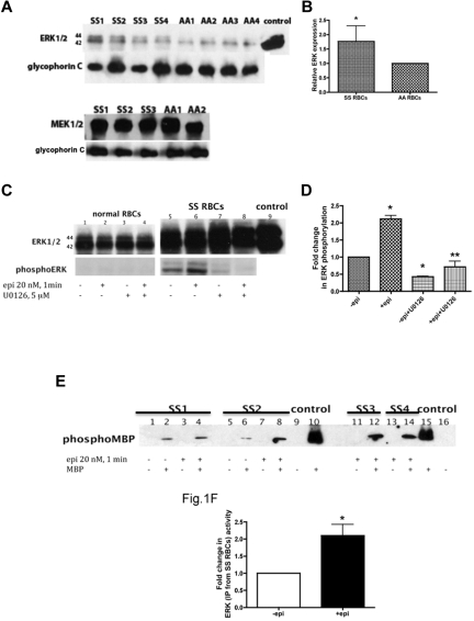
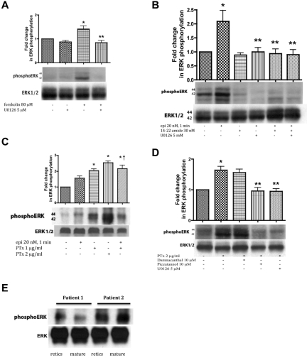
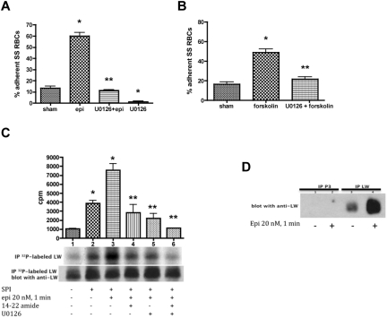
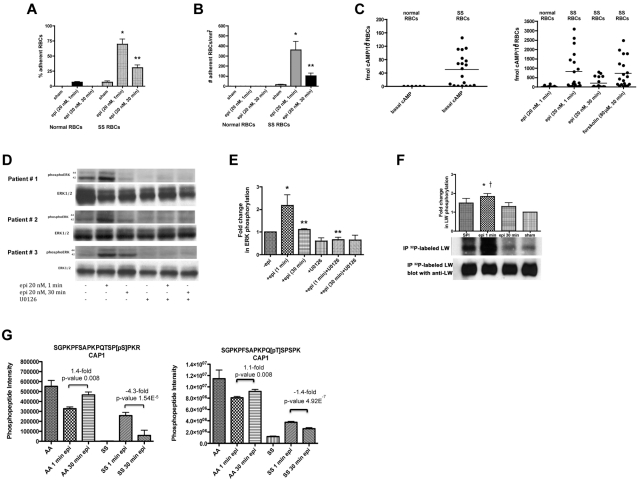
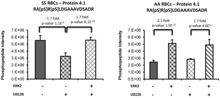
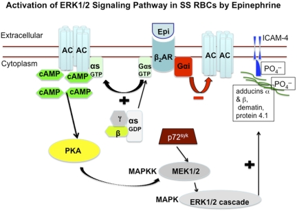
The core pathology of sickle cell disease (SCD) starts with the erythrocyte (RBC). Aberration in MAPK/ERK1/2 signaling, which can regulate cell adhesion, occurs in diverse pathologies. Because RBCs contain abundant ERK1/2, we predicted that ERK1/2 is functional in sickle (SS) RBCs and promotes adherence, a hallmark of SCD. ERK1/2 remained active in SS but not normal RBCs. β(2)-adrenergic receptor stimulation by epinephrine can enhance ERK1/2 activity only in SS RBCs via PKA- and tyrosine kinase p72(syk)-dependent pathways. ERK signaling is implicated in RBC ICAM-4 phosphorylation, promoting SS RBC adhesion to the endothelium. SS RBC adhesion and phosphorylation of both ERK and ICAM-4 all decreased with continued cell exposure to epinephrine, implying that activation of ICAM-4-mediated SS RBC adhesion is temporally associated with ERK1/2 activation. Furthermore, recombinant ERK2 phosphorylated α- and β-adducins and dematin at the ERK consensus motif. Cytoskeletal protein 4.1 also showed dynamic phosphorylation but not at the ERK consensus motif. These results demonstrate that ERK activation induces phosphorylation of cytoskeletal proteins and the adhesion molecule ICAM-4, promoting SS RBC adhesion to the endothelium. Thus, blocking RBC ERK1/2 activation, such as that promoted by catecholamine stress hormones, could ameliorate SCD pathophysiology.


Figures

Figure 1
Figure 1
ERK undergoes activation in SS but not normal RBCs. (A-B) Fifty micrograms of membrane protein ghosts (SS RBC ghosts, n = 4, lanes: SS1, SS2, SS3, and SS4; and normal RBC ghosts, n = 4, lanes: AA1, AA2, AA3, and AA4) were used per lane. Western blots of protein ghosts were stained with antibodies against ERK1/2, glycophorin C as a loading control, and MEK1/2 (n = 3 for SS RBC ghosts, lanes: SS1, SS2, and SS3; and n = 2 for normal RBC ghosts, lanes: AA1 and AA2). (A) ERK1/2 and MEK1/2 are highly expressed in both SS and normal RBCs and are bound to the RBC plasma membrane. (B) Quantitative analysis of the data (normalized according to glycophorin C expression) presented as relative ERK1/2 expression compared with normal RBCs (P < .05 for SS vs normal RBCs, n = 4 for each). (C-D) Normal RBCs (n = 3, lanes: 1, 2, 3, and 4) and SS RBCs (n = 3, lanes: 5, 6, 7, and 8) were sham-treated (lanes 1 and 5), incubated for 1 minute with 20nM epinephrine (epi; lanes 2 and 6), pretreated with the MEKI, U0126, followed by epi treatment (lanes 4 and 8), or treated with U0126 alone (lanes 3 and 7). Mouse 3T3/A31 fibroblast lysate was used as a positive control (lane 9). One hundred micrograms of SS and normal RBC ghost proteins were used per lane. Western blots were stained with antibodies against ERK and phosphoERK. (C) ERK1/2 is phosphorylated at baseline in SS RBCs and undergoes increased phosphorylation by epi stimulation. ERK in normal RBCs was not phosphorylated and completely failed to undergo increased phosphorylation after epi stimulation. (D) Quantitative analysis of the data is presented as fold change in ERK phosphorylation. *P < .01 compared with untreated cells. **P < .001 compared with epi-treated SS RBCs. (E-F) ERK immunoprecipitated from sham-treated (lanes: 1, 2, 5, and 6) and epi-treated (lanes: 3, 4, 7, 8, 11, 12, 13, and 14) SS RBCs was incubated without MBP (lanes: 1, 3, 5, 7, 11 and 13) or with MBP (lanes: 2, 4, 6, 8, 12, and 14) as a substrate for ERK, with equal protein amounts per assay condition. Commercial active recombinant human ERK2 was incubated without MBP (lanes: 9 and 16) or with MBP (lanes: 10 and 15) as negative and positive controls, respectively. (E) Immunoblots indicate that the activity of ERK is conserved and functional in SS RBCs and epi can intensify its activity. SS RBCs obtained from 4 different patients (SS1, SS2, SS3, and SS4) were tested. (F) Quantitative analysis of the data are presented as fold change in ERK phosphorylation (n = 4). *P = .0286 compared with nontreated cells.
Figure 2
Figure 2
ERK activation in SS RBCs involves the cAMP/PKA pathway and the tyrosine kinase p72syk and is sensitive to the effect of Gαi protein. SS RBCs (A-D), and reticulocyte-enriched and -depleted (mature) SS RBCs (E) were sham-treated, treated with forskolin (A), epi (B-C), PKAI, 14-22 amide, (B), or PTx (C-D) in the presence or absence of the MEKI U0126 (A,B,D), piceatannol (D) or damnacanthal (D). RBC proteins were blotted with antibodies against ERK and phosphoERK. Quantitative analysis of the blots is presented as fold change in ERK phosphorylation. (A-B) ERK undergoes phosphorylation via the cAMP/PKA pathway. (A) ERK undergoes increased phosphorylation after RBC incubation with forskolin, which is inhibited by U0126 (n = 3). *P < .05 compared with untreated cells. **P < .01 compared with forskolin-treated SS RBCs. (B) Phosphorylation of ERK is increased by epi, and this increase was abrogated by either 14-22 amide or U0126 (n = 3). *P < .01 compared with untreated cells. **P < .01 compared with epi-treated SS RBCs. (C) ERK phosphorylation in SS RBCs is enhanced by inactivation of the Gαi protein. PTx at either 1 or 2 μg/mL increased basal ERK phosphorylation (n = 9). *P < .001 compared with nontreated cells; †P < .05 compared with epi-treated SS RBCs. (D) The tyrosine kinase p72syk is implicated in ERK phosphorylation. PTx at 2 μg/mL up-regulated ERK phosphorylation, an effect that was blocked by piceatannol. Conversely, damnacanthal failed to block ERK phosphorylation induced by PTx (n = 3). *P < .01 compared with untreated cells. **P < .01 compared with PTx-treated SS RBCs. (E) ERK1/2 is phosphorylated at baseline in both reticulocyte-enriched and reticulocyte-depleted (mature) SS RBCs (n = 2).
Figure 3
Figure 3
ERK signaling modulates both SS RBC adhesion to endothelial cells and ICAM-4 phosphorylation. (A-B) Activation of ERK signaling up-regulates SS RBC adhesion to the endothelium. SS RBCs were sham-treated, stimulated with epi for 1 minute or forskolin, preincubated with U0126 followed by epi or forskolin, or treated with U0126 alone. Adhesion of SS RBCs to HUVECs was tested in intermittent flow condition assays. Results are presented as percent adherent SS RBCs at a shear stress of 2 dynes/cm2. Error bars show SEM of 4 different experiments. (A) *P < .001 compared with sham-treated; **P < .001 compared with epi-treated. (B) *P < .001 compared with sham-treated; **P < .001 compared with forskolin-treated. (C-D) The MEK/ERK signaling cascade is involved in ICAM-4 (LW) serine phosphorylation. (C) Inorganic 32P-radiolabeled intact SS RBCs were incubated in the absence (lane 1) or presence (lanes 2, 3, 4, 5, and 6) of serine/threonine protein phosphatase inhibitors (SPI), followed or not (lanes 1 and 2) by treatment with epi (lanes 3, 4, 5, and 6). In lanes 4, 5, and 6, SS RBCs were preincubated with SPI in presence of PKAI, MEKI, or PKAI + MEKI followed by epi treatment, respectively. The counts per minute (cpm) are representative of 3 different experiments, calculated by subtraction of cpm present in a lane (not shown) containing immunoprecipitates using immunoglobulin P3 from cpm obtained using anti-LW (ICAM-4) mAb for immunoprecipitation under each set of conditions indicated. *P < .05 and *P < .001 for SPI-treated and SPI + epi-treated vs sham-treated, respectively; **P < .001 compared with SPI + epi-treated SS RBCs. Total LW loaded in each lane was detected with the use of nitrocellulose membranes of phosphorylated LW blotted with anti-LW mAb. (D). SS RBCs were incubated without (lanes 1 and 3) or with epi (lanes 2 and 4). Lanes 1 and 2 were immunoprecipitated with P3. Lanes 3 and 4 were immunoprecipitated with anti-LW mAb; all lanes for panel D were immunostained with anti-LW mAb.
Figure 4
Figure 4
SS RBC adhesion is associated with the extent of ERK activation. (A-B) Adhesion of SS RBCs to endothelial cells is related to the duration of cell stimulation with epinephrine. Adhesion of RBCs to HUVECs was tested in both intermittent flow and flowing condition assays, and results are presented as precent adherent RBCs at a shear stress of 2 dynes/cm2 and number of adherent RBCs/mm2, respectively. Normal and SS RBCs were sham-treated, or stimulated with epi for 1 minute or 30 minutes. *P < .001 compared with sham-treated SS RBCs; **P < .001 compared with epi-treated SS RBCs. Error bars show SEM of 4 different experiments. (C) cAMP production in SS RBCs is associated with the time of cell stimulation with epinehprine. RBCs were treated with IBMX (for basal cAMP levels), followed or not with epi (for 1 minute or 30 minutes) or forskolin. The specific effect of epi and forskolin on cAMP accumulation was obtained by subtracting basal cAMP levels from the total cAMP levels. The basal cAMP production and specific amounts of cAMP because of epi or forskolin stimulation were normalized as fmol cAMP/108 RBCs. (D-E) ERK phosphorylation is dependent on the time of SS RBC exposure to epinephrine. SS RBCs were sham-treated or treated with epi for 1 or 30 minutes, U0126, or U0126 followed by epi for 1 or 30 minutes. Immunoblots of RBC proteins with antibodies against ERK and phosphoERK (D) and quantitative analysis of the data presented as fold change in ERK phosphorylation (E) are shown. ERK underwent increased phosphorylation after 1 minute exposure to epi, whereas phosphorylation decreased with longer (30 minutes) cell exposure to epi (n = 4). *P < .01 compared with nontreated cells; **P < .01 and **P < .001 for epi-treated for 30 minutes and U0126+epi-treated for 1 minute versus cells treated with epi for 1 minute, respectively (E). (F) Inorganic 32P radiolabeled intact SS RBCs were incubated in the presence (lanes 1, 2, and 3) or absence (lane 4) of SPI, followed (lanes 2 and 3) or not (lane 1) by treatment with epi for 1 minute (lane 2) or 30 minutes (lane 3). The cpm are representative of 3 different experiments, calculated by subtraction of cpm present in a lane (not shown) containing immunoprecipitates using immunoglobulin P3 from cpm obtained using anti-LW (ICAM-4) mAb for immunoprecipitation under each set of conditions indicated. *P < .01 compared with sham-treated; †P < .05 compared with SPI + epi (30 minutes)–treated SS RBCs. (G) Prolonged cell exposure to epinephrine negatively affects phosphorylation of adenylate cyclase-associated protein 1. RBC ghosts isolated from SS and normal RBCs treated with epi for 1 and 30 minutes were enriched in phosphopeptides and then subjected to a label-free quantitative phosphoproteomics analysis. Phosphorylation of both serine and threonine on CAP1 in SS RBCs decreased with increased time (1 minute vs 30 minutes) of cell exposure to epi, whereas an increase in the abundance of these phosphopeptides was observed in normal (AA) RBCs after 30 minutes exposure to epi. Each data point is an average of 3 analytical replicate measurements with error bars indicating SD.
Figure 5
Figure 5
Phosphorylation of protein 4.1 is induced via the ERK signaling pathway. Sham-treated or U0126-treated SS and normal (AA) RBCs ghosts coincubated with or without recombinant active ERK2 (ERK2) were enriched in phosphopeptides, followed by a label-free quantitative phosphoproteomics analysis. Treatment of SS RBCs with U0126 caused a significant decrease in doubly phosphorylated peptide within protein 4.1. The addition of ERK2 to the U0126-treated SS RBC ghosts increased the abundance of this phosphopeptide back to levels observed in untreated SS RBCs. The complementary trend for this phosphorylated peptide was also observed on the addition of ERK2 to AA RBCs sham-treated or U0126-treated.
Figure 6
Figure 6
Schematic depiction of proposed increased activation of ERK signaling pathway in SS RBCs. Epinephrine stimulates β2ARs on SS RBCs. β2ARs are prototypic G-coupled receptors whose signaling is largely mediated by activation of stimulatory GTP-binding proteins (Gs proteins), and inhibited by activation of Gαi protein. Activation of Gs proteins in turn activates AC, leading to generation of cAMP, and the subsequent activation of PKA. The activity of downstream signaling proteins, such as MEKs and ERKs is enhanced by PKA activation. The tyrosine kinase p72Syk acts synergistically with PKA to activate MEK/ERK cascade. Activation of ERK results in phosphorylation of the ERK consensus motif on the cytoskeletal proteins α- and β-adducins, dematin, and protein 4.1, albeit not at the ERK consensus motif. Phosphorylation of cytoskeletal proteins may result in cytoplasmic membrane protein conformational changes, which could render ICAM-4 accessible to phosphorylation.

Similar articles

Cited by

References

    1. Frenette PS, Atweh GF. Sickle cell disease: old discoveries, new concepts, and future promise. J Clin Invest. 2007;117(4):850–858. - PMC - PubMed
    1. Hebbel RP, Boogaerts MA, Eaton JW, Steinberg MH. Erythrocyte adherence to endothelium in sickle-cell anemia. A possible determinant of disease severity. N Engl J Med. 1980;302(18):992–995. - PubMed
    1. Mohandas N, Evans E. Sickle erythrocyte adherence to vascular endothelium. Morphologic correlates and the requirement for divalent cations and collagen-binding plasma proteins. J Clin Invest. 1985;76(4):1605–1612. - PMC - PubMed
    1. Zennadi R, Hines PC, De Castro LM, Cartron JP, Parise LV, Telen MJ. Epinephrine acts through erythroid signaling pathways to activate sickle cell adhesion to endothelium via LW-alphavbeta3 interactions. Blood. 2004;104(12):3774–3781. - PubMed
    1. Zennadi R, Chien A, Xu K, Batchvarova M, Telen MJ. Sickle red cells induce adhesion of lymphocytes and monocytes to endothelium. Blood. 2008;112(8):3474–3483. - PMC - PubMed

Publication types

MeSH terms

Substances