. 2012 Jan;139(1):75-83.
doi: 10.1242/dev.073692.

The muscle pattern of the Drosophila abdomen depends on a subdivision of the anterior compartment of each segment

Affiliations

The muscle pattern of the Drosophila abdomen depends on a subdivision of the anterior compartment of each segment

Joanna Krzemien et al. Development. 2012 Jan.

Abstract

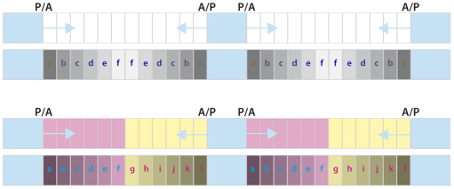
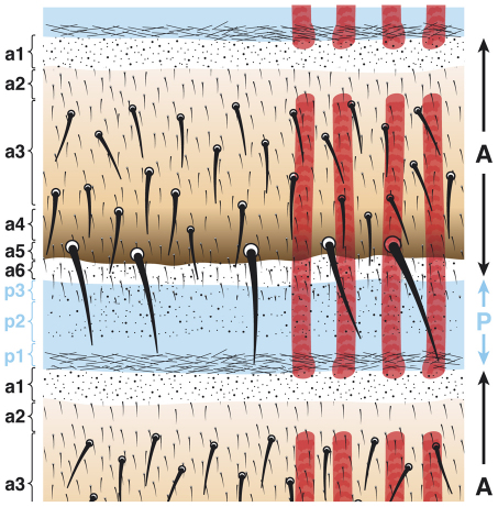
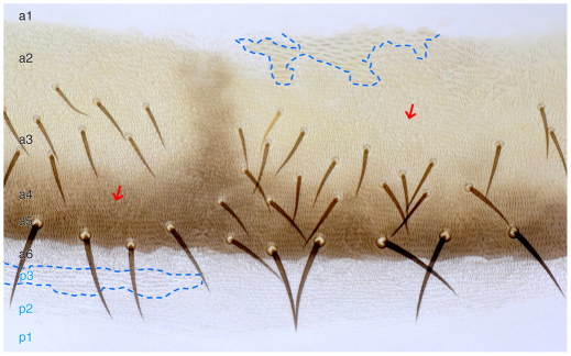
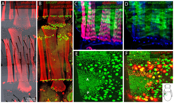
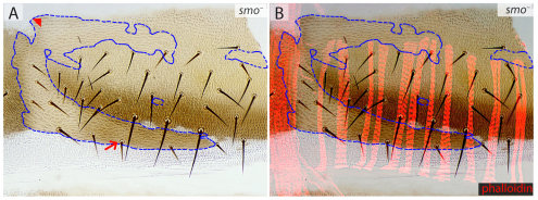
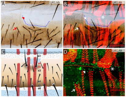
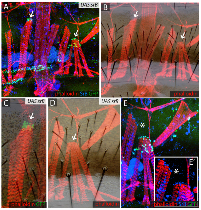
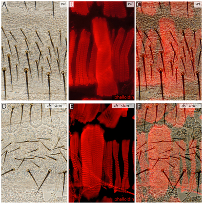
In the past, segments were defined by landmarks such as muscle attachments, notably by Snodgrass, the king of insect anatomists. Here, we show how an objective definition of a segment, based on developmental compartments, can help explain the dorsal abdomen of adult Drosophila. The anterior (A) compartment of each segment is subdivided into two domains of cells, each responding differently to Hedgehog. The anterior of these domains is non-neurogenic and clones lacking Notch develop normally; this domain can express stripe and form muscle attachments. The posterior domain is neurogenic and clones lacking Notch do not form cuticle; this domain is unable to express stripe or form muscle attachments. The posterior (P) compartment does not form muscle attachments. Our in vivo films indicate that early in the pupa the anterior domain of the A compartment expresses stripe in a narrowing zone that attracts the extending myotubes and resolves into the attachment sites for the dorsal abdominal muscles. We map the tendon cells precisely and show that all are confined to the anterior domain of A. It follows that the dorsal abdominal muscles are intersegmental, spanning from one anterior domain to the next. This view is tested and supported by clones that change cell identity or express stripe ectopically. It seems that growing myotubes originate in posterior A and extend forwards and backwards until they encounter and attach to anterior A cells. The dorsal adult muscles are polarised in the anteroposterior axis: we disprove the hypothesis that muscle orientation depends on genes that define planar cell polarity in the epidermis.


Figures

Fig. 1.
Fig. 1.
Compartments and morphogens in pattern formation. The insect body plan consists of a chain of metameres divided into anterior (A) and posterior (P) compartments. Hedgehog (Hh) is produced by all the P cells and spreads forwards and backwards into the adjacent A compartments, forming concentration gradients (blue arrows) that pattern the A cells. One scenario is shown at the top: all the A cells might respond to Hh alike giving a reflexed pattern of cell types (a-f-a). However, this pattern is not observed and instead there is a single sequence of cuticle types (a-l); our explanation depends on there being two types of A cells (shown in magenta and yellow) each responding differently to Hh (Struhl et al., 1997b).
Fig. 2.
Fig. 2.
The cuticle of an adult abdominal segment. The epidermis of the Drosophila adult abdomen derives from nests of histoblasts that are set aside in the embryo and remain quiescent throughout the larval stages. In each adult segment, nine different cuticle types can be distinguished by means of surface structure, pigmentation and bristles. The A compartment is subdivided into a smaller anterior domain composed of a1 and a2 cuticle and a larger posterior domain formed by a3-a6 cuticle. These two regions are distinguished because their cells respond to Hh signalling differently (see Fig. 1) (Struhl et al., 1997b) and only the posterior domain requires N (see Fig. 3). Note, it is difficult to place the border precisely between a2 and a3; a2 was distinguished from a3 only by the absence of bristles (Struhl et al., 1997b). The bristles move during development (García-Bellido and Merriam, 1971) and therefore are not a reliable marker of the provenance of the epidermal cells around them. The dorsal abdominal muscles of one side are shown in red. Anterior is at the top and the posterior is at the bottom.
Fig. 3.
Fig. 3.
Differential requirement for N in the A compartment. Marked N clones in the dorsal cuticle (outlined with blue dashed lines). Clones arising in the posterior domain of A (a3-a6) do not make cuticle. We now correct our earlier report (Lawrence et al., 2002) when a6 was not included in that posterior zone: of 24 N clones found close to and on either side of the a6/P border in 11 abdomens only two were found in a6. These two exceptions were near the midline where N clones sometimes can be seen, even in a3 territory. Apart from these occasional exceptions, a3 epidermis is all unmarked (N+ cells) but has areas that lack bristles (indicated by arrows). These bald patches could be due to N clones that made neurons there and inhibited nearby bristles. We stained for neurons and found clusters of neurons of different sizes underlying the bald patches of cuticle. Of four bald patches, varying in size from one-third to most of a hemisegment, we saw underneath the cuticle five, eight, eight and ten clusters of neurons. No such clusters were seen in the normal territory flanking the bald patches. Perhaps, as in the notum of Drosophila, each N cell forms neurons that emit a strong Delta signal, inhibiting the formation of N+ bristles nearby (Heitzler and Simpson, 1991). However, clones form normally in the anterior domain of the A compartment and also in the P compartment and this image shows both.
Fig. 4.
Fig. 4.
Muscle attachments in the dorsal abdominal epidermis. (A,B) Both anterior and posterior attachment sites of the Drosophila larval persistent muscles (larger muscles) are located in a2 cuticle, whereas the anterior attachment sites for adult muscles (the smaller muscles) are located near to the boundary between a2 and a3 (see Fig. 2). The nuclei of tendon cells are labelled red with sr.Gal4 UAS.RFP, the muscular attachments are marked in green with Ilk::GFP and the muscles are marked in red with phalloidin. (C,D) Posterior attachment sites of adult muscles (tendon nuclei, marked blue with anti-SrB antibody) are located in the first rows of the A compartment, just behind the cells of the en-expressing P compartment (patchy green). C and D show that the tendon nuclei and the P cells are adjacent but do not overlap. (E,F) Dorsolateral view of a segment ∼21 hours after puparium formation. (E) Two histoblast nests: the bigger anterior (A) and the smaller posterior (P) fuse to form the adult epidermis of one segment. All nuclei, larval (large) and adult (small), express H2A::GFP. (F) A merge of red and green channels shows that the sr-expressing adult cells (red) are located at the front of the anterior nest. The diagram shows the position of the analysed histoblast nests in the pupa.
Fig. 5.
Fig. 5.
smo clones in the cuticle. (A,B) smo clones (marked with y and stc, encircled by blue dashed lines). The identity of the smo cells in the posterior A compartment is changed into a3 (small bristles, arrow) and in the anterior A compartment a1 is transformed into a2 (arrowhead) We believe that the muscles (red, B) and their attachment sites are not affected by these transformations because a3 and P are incompetent to form attachments, and a1 and a2 are competent and equivalent.
Fig. 6.
Fig. 6.
UAS.hh clones change the identities of the epidermal cells as well as the positions of muscle attachments. (A-C) UAS.hh-expressing clone (boundary estimated with a blue dashed line) located in a2/a3 area, transforms its cells towards P identity and as Hh spreads from the clone into surrounding A territory it changes the identity and orientation of neighbouring cells, replacing a2/a3 and a3 with a6-a4 (Struhl et al., 1997b) as shown schematically in C. The muscles underlying the transformed cuticle do not contact the newly formed a4-a6 cuticle and instead attach to a1 cuticle that is still present (asterisk). Note that muscles from the preceding segment attach correctly even in the neighbourhood of a UAS.hh clone, presumably because a1 is not changed by the clone. (D) Detail showing the muscle attachments and apparently normal tendon cells (green, marked with anti-SrB). Asterisks, arrows and arrowheads indicate the corresponding points in the three figures.
Fig. 7.
Fig. 7.
UAS.sr clones attract muscles and divert them from their usual points of attachment. (A,B) UAS.srB clones (marked with GFP, green) attract the muscles (stained with phalloidin, red). All tendon nuclei are marked with anti-SrB antibody (blue). Some muscles attach to ectopic sites where sr is expressed (arrows). (C-E′) Details of clones shown in A and B. Note the altered cuticle in one sr-expressing clone (arrow, C) while another clone can make maimed bristles (below arrow, D). (E) The same region stained for anti-SrB antibody (blue) and GFP for UAS.srB (green); (E′) A detail of E lacking the green channel showing that both the endogenous and ectopic tendons express sr at similar levels. It appears that myotubes ignore their usual attachment sites and prefer to form ectopic contacts with the clones, either because the ectopic sr expression starts earlier or is more persistent; if the latter, this could be due to autoregulation of sr (Vorbruggen and Jackle, 1997). Asterisk in E,E′ indicates the same location.
Fig. 8.
Fig. 8.
In ds stan mutant flies, muscle orientation is not affected. The bristles and hairs are oriented by the Stan and Ds systems, the twin mechanisms of planar cell polarity (PCP) (Casal et al., 2006; Lawrence et al., 2007). (A-C) Wild-type dorsal epidermis and the underlying muscles. (D-F) ds stan flies have disoriented hairs and bristles but the identity of the epidermal cells is not changed. Muscle orientation is not affected by lack of organised polarity in the cuticle. Muscles are stained with phalloidin.

Similar articles

Cited by

References

    1. Akam M. (1987). The molecular basis for metameric pattern in the Drosophila embryo. Development 101, 1–22 - PubMed
    1. Bate M. (1990). The embryonic development of larval muscles in Drosophila. Development 110, 791–804 - PubMed
    1. Bate M., Rushton E., Currie D. A. (1991). Cells with persistent twist expression are the embryonic precursors of adult muscles in Drosophila. Development 113, 79–89 - PubMed
    1. Becker S., Pasca G., Strumpf D., Min L., Volk T. (1997). Reciprocal signaling between Drosophila epidermal muscle attachment cells and their corresponding muscles. Development 124, 2615–2622 - PubMed
    1. Bischoff M., Cseresnyes Z. (2009). Cell rearrangements, cell divisions and cell death in a migrating epithelial sheet in the abdomen of Drosophila. Development 136, 2403–2411 - PMC - PubMed

Publication types

MeSH terms

LinkOut - more resources