Review
. 2011 Nov 21;17(43):4757-71.
doi: 10.3748/wjg.v17.i43.4757.

Spectrum of mucin-producing neoplastic conditions of the abdomen and pelvis: cross-sectional imaging evaluation

Affiliations
Review

Spectrum of mucin-producing neoplastic conditions of the abdomen and pelvis: cross-sectional imaging evaluation

Nam Kyung Lee et al. World J Gastroenterol. .

Abstract

Various mucin-producing neoplasms originate in different abdominal and pelvic organs. Mucinous neoplasms differ from non-mucinous neoplasms because of the differences in clinical outcome and imaging appearance. Mucinous carcinoma, in which at least 50% of the tumor is composed of large pools of extracellular mucin and columns of malignant cells, is associated with a worse prognosis. Signet ring cell carcinoma is characterized by large intracytoplasmic mucin vacuoles that expand in the malignant cells with the nucleus displaced to the periphery. Its prognosis is also generally poor. In contrast, intraductal papillary mucinous neoplasm of the bile duct and pancreas, which is characterized by proliferation of ductal epithelium and variable mucin production, has a better prognosis than other malignancies in the pancreaticobiliary tree. Imaging modalities play a critical role in differentiating mucinous from non-mucinous neoplasms. Due to high water content, mucin has a similar appearance to water on ultrasound (US), computed tomography (CT), and magnetic resonance imaging, except when thick and proteinaceous, and then it tends to be hypoechoic with fine internal echoes or have complex echogenicity on US, hyperdense on CT, and hyperintense on T1- and hypointense on T2-weighted images, compared to water. Therefore, knowledge of characteristic mucin imaging features is helpful to diagnose various mucin-producing neoplastic conditions and to facilitate appropriate treatment.

Keywords: Abdomen and pelvis; Computed tomography; Magnetic resonance; Mucin; Neoplasm; Ultrasound.


Figures

Figure 1
Figure 1
Mucinous cystadenoma of the pancreas in a 40-year-old woman. A: Endoscopic ultrasound image shows a complex multiloculated cystic mass in the pancreatic tail. Note a hyperechoic locule (asterisk) within the cystic tumor; B: Contrast-enhanced CT image shows a well-circumscribed multiloculated mass in the tail of the pancreas with enhancement of thin internal septa and the peripheral wall. Note a highly attenuated locule (arrow) within the cystic tumor. CT: Computed tomography.
Figure 2
Figure 2
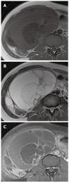
Mucinous cystadenocarcinoma of the pancreas in a 44-year-old woman. A, B: Non-enhanced (A) and contrast-enhanced (B) CT images show a well-circumscribed multi-septated cystic mass with enhancing soft tissue components (arrowheads) in the tail of the pancreas; C: T2-weighted MR image shows a large multilocular cystic mass with high signal intensity and peripheral soft tissue components with low signal intensity (arrowheads) in the pancreatic tail. CT: Computed tomography; MR: Magnetic resonance.
Figure 3
Figure 3
Biliary cystadenoma in the liver of a 55-year-old woman. A: Ultrasound image shows a multiseptated cystic mass in the liver; B: Contrast-enhanced CT image shows a well-circumscribed cystic liver mass, which has similar attenuation to that of hepatic cysts. Note the internal septa with nodular thickening (arrowhead); C, D: T1- (C) and T2-weighted (D) MR images show a multiseptated cystic mass with hyperintensity. CT: Computed tomography; MR: Magnetic resonance.
Figure 4
Figure 4
Coexistence of a mucinous cystadenoma of the appendix and ovary in a 63-year-old woman. A: Coronal T2-weighted MR image shows a well-defined cystic mass with hyperintensity in the appendix (arrow) and a multiloculated cystic mass in the left ovary (asterisk); B, C: Contrast-enhanced CT images show low-attenuation mucinous deposits in the peritoneal cavity and scalloping of the liver margin (arrowheads), suggestive of pseudomyxoma peritonei. CT: Computed tomography; MR: Magnetic resonance.
Figure 5
Figure 5
Mucinous borderline tumor of the right ovary in a 41-year-old woman. A, B: T1- (A) and T2-weighted (B) MR images show a cystic mass with large numbers of loculi with varying signal intensity in the right ovary; C: Contrast-enhanced fat-saturated T1-weighted MR image shows the enhancement of multiple internal septa and wall in the cystic mass. MR: Magnetic resonance.
Figure 6
Figure 6
Mucinous cystadenocarcinoma of the left ovary in a 47-year-old woman. A: Sagittal T2-weighted MR image shows a huge multilocular ovarian cystic mass with multiple hypointense solid components (arrowheads); B: Sagittal contrast-enhanced T1-weighted MR image shows an ovarian cystic mass with enhancement of internal septa and solid components (arrowheads). MR: Magnetic resonance.
Figure 7
Figure 7
Mucocele of the appendix in a 54-year-old man. Ultrasound image shows an anechoic round mass in the appendix with echogenic layers (arrowhead) (the so-called “onion-skin” sign).
Figure 8
Figure 8
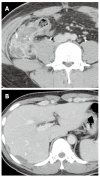
Mucinous cystadenocarcinoma of the appendix in a 26-year-old man. A: Contrast-enhanced CT image shows a complex hypoattenuating mass with enhancing solid portions and punctate calcifications in the appendix (arrow); B: Contrast-enhanced CT image of the upper abdomen shows loculations of fluid scalloping on the liver surface (arrowheads), providing evidence of a mass effect. CT: Computed tomography.
Figure 9
Figure 9
Intraductal papillary mucinous neoplasm of the pancreas in an 80-year-old woman. A: Coronal T2-weighted RARE MR image shows cord-like hypointense mucin in the dilated main pancreatic duct (arrow). Note multiple stones in the dilated common bile duct; B: Axial contrast-enhanced fat-saturated T1-weighted MR image demonstrates a fistula (asterisk) between the dilated main pancreatic duct and the stomach. Note mural nodules (arrowheads) within the dilated main pancreatic duct, which strongly suggest a malignant Intraductal papillary mucinous neoplasm; C: Endoscopy image reveals mucin leaking from the papilla. RARE: True rapid acquisition with relaxation enhancement; MR: Magnetic resonance.
Figure 10
Figure 10
Intraductal papillary mucinous neoplasm of the bile duct in a 58-year-old woman. A: Contrast-enhanced CT image shows severe dilatation of the left intrahepatic ducts without visible intraductal mass. Note a small stone in the dilated intrahepatic duct (arrowhead); B: Coronal T2-weighted RARE MR image shows dilatation of the intrahepatic and extrahepatic ducts. Note the hypointense intraductal mass (arrow) in the left intrahepatic duct; C: Endoscopic retrograde cholangiogram image shows a filling defect (arrow) within the marked dilated extrahepatic bile duct, due to mucin; D: Coronal gadoxetic acid-enhanced T1-weighted MR image obtained at 60 min post-injection shows contrast-filled bile duct with an elongated, low signal intensity lesion (asterisk), which represents mucin; E: Endoscopy image reveals mucin leaking from the papilla. CT: Computed tomography; RARE: True rapid acquisition with relaxation enhancement; MR: Magnetic resonance.
Figure 11
Figure 11
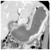
Signet ring cell carcinoma of the stomach in a 45-year-old woman. Coronal reformatted contrast-enhanced CT image shows diffuse gastric wall thickening with strong enhancement (arrowhead) along the lesser and greater curvature of the stomach. CT: Computed tomography.
Figure 12
Figure 12
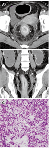
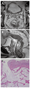
Signet ring cell carcinoma of the rectum in a 42-year-old man. A, B: Axial (A) and coronal reformatted (B) contrast-enhanced CT images show concentric wall thickening with malignant target sign (arrowheads in A, arrow in B) in the rectum; C: Photomicrograph image (original magnification, × 200; HE stain) shows multiple signet ring cells. CT: Computed tomography; HE: Hematoxylin and eosin.
Figure 13
Figure 13
Mucinous carcinoma of the stomach in a 62-year-old woman. A: Contrast-enhanced CT image shows a large mass lesion in the gastric cardia containing abundant mucin pools (asterisk) and enhanced solid portions (arrow); B: Endoscopy image shows jellylike mucin leaking from the gastric mass. CT: Computed tomography.
Figure 14
Figure 14
Mucinous adenocarcinoma of the cecum in a 69-year-old man. Contrast-enhanced CT image shows a huge eccentric hypoattenuating mass with poor enhancement of the solid portion of the cecum (asterisk). The mass has invaded the retroperitoneum (arrow). CT: Computed tomography.
Figure 15
Figure 15
Mucinous adenocarcinoma of the rectum in a 63-year-old man. A, B: Axial (A) and coronal (B) T2-weighted MR images show an area of hyperintense mucin pools (arrowhead) in the rectal mass; C: Photomicrograph image (original magnification, × 40; HE stain) shows large pools of extracellular mucin and tumor cells. MR: Magnetic resonance; HE: Hematoxylin and eosin.
Figure 16
Figure 16
Perianal mucinous adenocarcinoma in a 41-year-old man. A: Sagittal T2-weighted MR image shows a hyperintense mass (arrowheads) in the perianal area; B: Sagittal contrast-enhanced T1-weighted MR image shows mesh-like internal enhancement (arrowheads) within the mass; C: Photomicrograph image (original magnification, × 100; HE stain) shows large pools of extracellular mucin and tumor cells. MR: Magnetic resonance; HE: Hematoxylin and eosin.
Figure 17
Figure 17
Intraperitoneal spread of mucinous adenocarcinoma of the urachus (also called peritoneal mucinous carcinomatosis) in a 57-year-old man. A: Contrast-enhanced CT image shows a midline supravesical mass with heterogeneous attenuation. Within the mass are scattered low-attenuation areas (arrowheads), which represent mucin; B: Contrast-enhanced CT image of the upper abdomen shows low-attenuation mucinous ascites scalloping the liver margin. Note the right pleural effusion (asterisk) and diffuse nodular thickening of the peritoneum (arrowheads). CT: Computed tomography; MR: Magnetic resonance.
Figure 18
Figure 18
Adenoma malignum of the cervix in a 30-year-old woman with Peutz-Jeghers syndrome. A: Axial T2-weighted MR image shows a multicystic lesion with a solid component (arrowheads) in the uterine cervix; B: Contrast-enhanced CT image shows multiple polyps (arrows) in the duodenum and small bowel causing intussusceptions. CT: Computed tomography; MR: Magnetic resonance.
Figure 19
Figure 19
Mucinous carcinoma of the gallbladder in a 77-year-old woman. A: Contrast-enhanced CT image shows localized wall thickening (arrow) in the body of the gallbladder containing a suspicious mucin pool; B: Contrast-enhanced CT image inferior to (A) shows a punctate calcification and localized wall thickening (arrow) in a mildly distended gallbladder. CT: Computed tomography.

Similar articles

Cited by

References

    1. Zhang M, Zhu GY, Zhang HF, Gao HY, Han XF, Xue YW. Clinicopathologic characteristics and prognosis of mucinous gastric carcinoma. J Surg Oncol. 2010;102:64–67. - PubMed
    1. Consorti F, Lorenzotti A, Midiri G, Di Paola M. Prognostic significance of mucinous carcinoma of colon and rectum: a prospective case-control study. J Surg Oncol. 2000;73:70–74. - PubMed
    1. Maehara Y, Sakaguchi Y, Moriguchi S, Orita H, Korenaga D, Kohnoe S, Sugimachi K. Signet ring cell carcinoma of the stomach. Cancer. 1992;69:1645–1650. - PubMed
    1. Tung SY, Wu CS, Chen PC. Primary signet ring cell carcinoma of colorectum: an age- and sex-matched controlled study. Am J Gastroenterol. 1996;91:2195–2199. - PubMed
    1. Lim JH, Lee G, Oh YL. Radiologic spectrum of intraductal papillary mucinous tumor of the pancreas. Radiographics. 2001;21:323–337; discussion 337-340. - PubMed

Publication types

MeSH terms