

Benign cephalic histiocytosis: a case report
- PMID: 22148022
- PMCID: PMC3229948
- DOI: 10.5021/ad.2011.23.4.508
Benign cephalic histiocytosis: a case report
Abstract
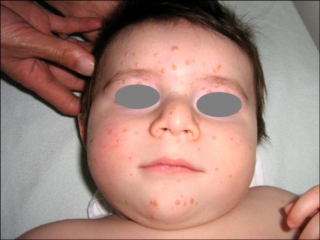
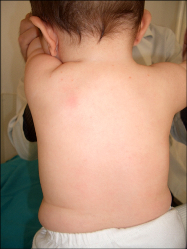
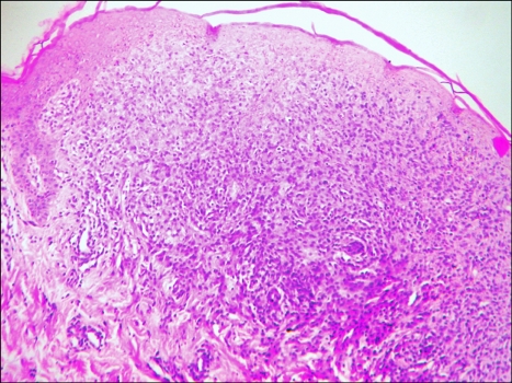
Histiocytic skin disorders are usually classified as either Langerhans' cell histiocytosis (LCH) or non LCH, based on the pathology. Benign cephalic histiocytosis (BCH) is a rare type of non-Langerhans histiocytitic disorder and is characterized by self-healing multiple small eruptions of yellow to red-brown papules on the face and upper trunk. Histologic features of this disorder show dermal proliferation of histiocytes that have intracytoplasmic comma-shaped bodies, coated vesicles and desmosome-like structures. In this study, we report on a 7-month-old boy who contained small yellow-red papules on his face that spread to his upper trunk. The clinical and histologic features in this patient were consistent with BCH.
Keywords: Benign cephalic histiocytosis; Generalized eruptive histiocytoma; Infant; Juvenile xanthogranuloma; Non-Langerhans-Cell.
Figures

Similar articles
-
Benign cephalic histiocytosis: a case report and review.J Am Acad Dermatol. 2002 Dec;47(6):908-13. doi: 10.1067/mjd.2002.124602. J Am Acad Dermatol. 2002. PMID: 12451377 Review.
-
Benign cephalic histiocytosis: case report and review of the literature.Pediatr Dermatol. 2014 Sep-Oct;31(5):547-50. doi: 10.1111/pde.12135. Epub 2013 Apr 3. Pediatr Dermatol. 2014. PMID: 23551579 Review.
-
Benign cephalic histiocytosis progressing into juvenile xanthogranuloma: a non-Langerhans cell histiocytosis transforming under the influence of a virus?Am J Dermatopathol. 2000 Feb;22(1):70-4. doi: 10.1097/00000372-200002000-00014. Am J Dermatopathol. 2000. PMID: 10698221
-
Indeterminate cell histiocytosis that presented clinically as benign cephalic histiocytosis.Dermatol Online J. 2014 Dec 16;20(12):13030/qt23d5k96k. Dermatol Online J. 2014. PMID: 25526330
-
Multiple eruptive cephalic histiocytomas in a case of T-cell lymphoma. A xanthomatous stage of benign cephalic histiocytosis in an adult patient?Am J Dermatopathol. 1993 Dec;15(6):581-6. doi: 10.1097/00000372-199312000-00013. Am J Dermatopathol. 1993. PMID: 8311192
Cited by
-
Benign cephalic histiocytosis in a 2-year-old boy with an inconspicuous clinical presentation at onset.Clin Case Rep. 2023 Oct 10;11(10):e8043. doi: 10.1002/ccr3.8043. eCollection 2023 Oct. Clin Case Rep. 2023. PMID: 37830061 Free PMC article.
-
Successful Treatment of Non-Langerhans Cell Histiocytosis With Topical Rapamycin in Two Pediatric Cases.Clin Cosmet Investig Dermatol. 2022 Aug 6;15:1575-1582. doi: 10.2147/CCID.S375995. eCollection 2022. Clin Cosmet Investig Dermatol. 2022. PMID: 35967913 Free PMC article.
-
Unraveling cutaneous histiocytosis: insights into histology, pathogenesis, diagnosis, and treatment pitfalls.Front Med (Lausanne). 2025 Jun 20;12:1585815. doi: 10.3389/fmed.2025.1585815. eCollection 2025. Front Med (Lausanne). 2025. PMID: 40620437 Free PMC article. Review.
-
Benign cephalic histiocytosis.Indian Dermatol Online J. 2013 Oct;4(4):300-1. doi: 10.4103/2229-5178.120646. Indian Dermatol Online J. 2013. PMID: 24350010 Free PMC article.
References
-
- Gianotti F, Caputo R, Ermacora E. Singular "infantile histiocytosis with cells with intracytoplasmic vermiform particles". Bull Soc Fr Dermatol Syphiligr. 1971;78:232–233. - PubMed
-
- Jih DM, Salcedo SL, Jaworsky C. Benign cephalic histiocytosis: a case report and review. J Am Acad Dermatol. 2002;47:908–913. - PubMed
-
- Zelger BW, Sidoroff A, Orchard G, Cerio R. Non-Langerhans cell histiocytoses. A new unifying concept. Am J Dermatopathol. 1996;18:490–504. - PubMed
-
- Dadzie O, Hopster D, Cerio R, Wakeel R. Benign cephalic histiocytosis in a British-African child. Pediatr Dermatol. 2005;22:444–446. - PubMed
-
- Hasegawa S, Deguchi M, Chiba-Okada S, Aiba S. Japanese case of benign cephalic histiocytosis. J Dermatol. 2009;36:69–71. - PubMed
Publication types
LinkOut - more resources
Full Text Sources
