

Primary localized cutaneous nodular amyloidosis following local trauma
- PMID: 22148024
- PMCID: PMC3229950
- DOI: 10.5021/ad.2011.23.4.515
Primary localized cutaneous nodular amyloidosis following local trauma
Abstract
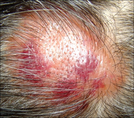
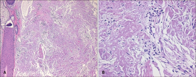
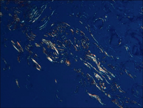
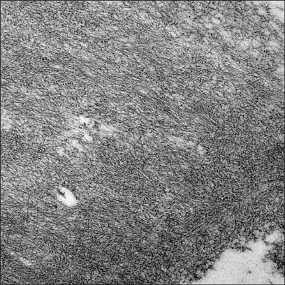
Primary localized cutaneous nodular amyloidosis (nodular amyloidosis) is a rare and distinct type of amyloidosis, in which amyloid L deposition is limited to the skin and typically manifested as a tumefactive nodule on the acral sites. However, the definite cause of nodular amyloidosis is still unknown. Although it is relatively well known that the amyloid deposits in nodular amyloidosis originate from immunoglobulin light chains secreted by local plasma cells, traumatic injury to the skin has rarely been recognized as a triggering factor of nodular amyloidosis. Herein, we present a case of a 50-year-old male patient with primary localized cutaneous nodular amyloidosis, which occurred after local trauma, and discuss the relationship between traumatic damage and dermal amyloid L deposition.
Keywords: Primary localized cutaneous nodular amyloidosis; Trauma.
Figures
References
-
- Breathnach SM. Amyloid and amyloidosis. J Am Acad Dermatol. 1988;18:1–16. - PubMed
-
- James WD, Berger TG, Elston DM. Andrew's diseases of the skin: clinical dermatology. 10th ed. Philadelphia: Saunders Elsevier; 2006. pp. 519–522.
-
- Elder DE, Elenitsas R, Johnson BL, Jr, Murphy GF, Xu X. Lever's histopathology of the skin. 10th ed. Philadelphia: Lippincott Williams & Wilkins; 2009. pp. 870–876.
-
- Kalajian AH, Waldman M, Knable AL. Nodular primary localized cutaneous amyloidosis after trauma: a case report and discussion of the rate of progression to systemic amyloidosis. J Am Acad Dermatol. 2007;57(2 Suppl):S26–S29. - PubMed
Publication types
LinkOut - more resources
Full Text Sources
