

Case Reports
doi: 10.5021/ad.2011.23.4.536.
Epub 2011 Nov 3.
Squamous Cell Carcinoma Developing within Lesions of Disseminated Superficial Actinic Porokeratosis
Affiliations
- PMID: 22148029
- PMCID: PMC3229955
- DOI: 10.5021/ad.2011.23.4.536
Item in Clipboard
Case Reports
Squamous Cell Carcinoma Developing within Lesions of Disseminated Superficial Actinic Porokeratosis
Ann Dermatol.
2011 Nov.
Abstract
Disseminated superficial actinic porokeratosis (DSAP) consists of multiple annular, hyperkeratotic lesions that have a bilateral distribution on sun-exposed areas, particularly the extremities. DSAPs have a wider distribution than porokeratosis of Mibelli and usually develop during the 3rd or 4th decade of life. Squamous cell carcinoma that arises in the classical type of porokeratosis of Mibelli is well-documented, but there are only a few reports of squamous cell carcinoma in DSAP. Here, we describe a 62-year-old man with DSAP who developed squamous cell carcinoma on his right forearm.
Keywords: Disseminated superficial actinic porokeratosis; Squamous cell carcinoma.
Figures
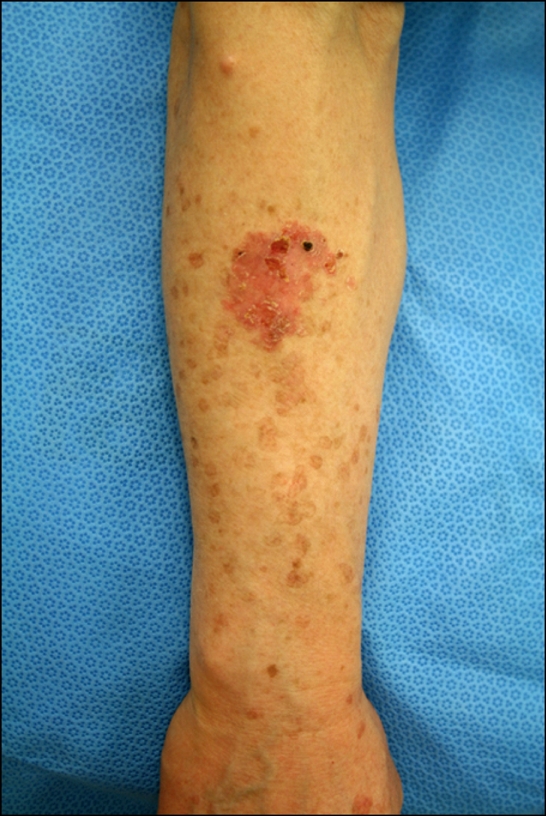
An irregular, marginated, erythematous plaque and multiple, brown, atrophic macules surrounded by well-demarcated, raised ridges on the right forearm.
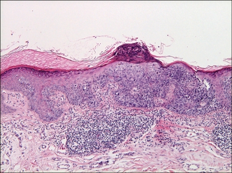
Biopsy specimen obtained from the erythematous plaque on the right arm. In epidermis, acanthosis and dysregulated keratinocytes with hyperchromatic, atypical nuclei are observed. A cornoid lamella composed of a column of parakeratosis is seen in the lesion of the squamous cell carcinoma (H&E, ×100).
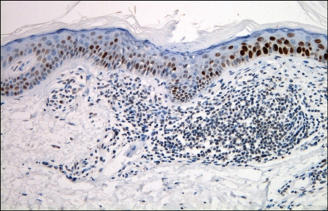
Overexpression of p53 in the epidermis of a disseminated superficial actinic porokeratosis lesion. A column of parakeratosis with underlying hypogranulosis is observed. Perivascular lymphocytic infiltrations are localized beneath the cornoid lamella (p53 immunohistochemical stain, ×200).
Similar articles
-
Disseminated Superficial Actinic Porokeratosis (DSAP): A Case Report Highlighting the Clinical, Dermatoscopic, and Pathology Features of the Condition.Cureus. 2022 Jul 16;14(7):e26923. doi: 10.7759/cureus.26923. eCollection 2022 Jul. Cureus. 2022. PMID: 35983404 Free PMC article.
-
Disseminated Superficial Actinic Porokeratosis.2025 Apr 6. In: StatPearls [Internet]. Treasure Island (FL): StatPearls Publishing; 2025 Jan–. 2025 Apr 6. In: StatPearls [Internet]. Treasure Island (FL): StatPearls Publishing; 2025 Jan–. PMID: 29083728 Free Books & Documents.
-
Disseminated superficial actinic porokeratosis co-existing with linear and verrucous porokeratosis in an elderly woman: Update on the genetics and clinical expression of porokeratosis.J Am Acad Dermatol. 2010 Nov;63(5):886-91. doi: 10.1016/j.jaad.2009.07.038. Epub 2010 May 6. J Am Acad Dermatol. 2010. PMID: 20451293 Review.
-
Squamous cell carcinoma arising from a lesion of disseminated superficial actinic porokeratosis.Clin Exp Dermatol. 1991 Nov;16(6):460-2. doi: 10.1111/j.1365-2230.1991.tb01237.x. Clin Exp Dermatol. 1991. PMID: 1806324 Review.
-
[Squamous cell carcinoma and disseminated superficial actinic porokeratosis].Ann Dermatol Venereol. 1994;121(1):50-2. Ann Dermatol Venereol. 1994. PMID: 8092731 French.
Cited by
-
Disseminated Superficial Actinic Porokeratosis in a Mother and Daughter: A Case Report.Cureus. 2025 Mar 18;17(3):e80796. doi: 10.7759/cureus.80796. eCollection 2025 Mar. Cureus. 2025. PMID: 40255783 Free PMC article.
-
A Case of Squamous Cell Carcinoma Arising in Disseminated Superficial Porokeratosis.Clin Cosmet Investig Dermatol. 2024 May 29;17:1259-1263. doi: 10.2147/CCID.S463569. eCollection 2024. Clin Cosmet Investig Dermatol. 2024. PMID: 38827628 Free PMC article.
-
Disseminated superficial actinic porokeratosis on the face treated with imiquimod 5% cream.Case Rep Dermatol. 2013 Oct 9;5(3):283-9. doi: 10.1159/000355180. eCollection 2013. Case Rep Dermatol. 2013. PMID: 24403891 Free PMC article.
References
-
- Shumack SP, Commens CA. Disseminated superficial actinic porokeratosis: a clinical study. J Am Acad Dermatol. 1989;20:1015–1022. - PubMed
-
- Schwarz T, Seiser A, Gschnait F. Disseminated superficial "actinic" porokeratosis. J Am Acad Dermatol. 1984;11:724–730. - PubMed
-
- Yang HY, Nam TS, Kim YT, Kim JH. A case of squamous cell carcinoma and Bowen's disease associated with superficial disseminated porokeratosis. Ann Dermatol. 1990;2:31–34.
-
- James WD, Rodman OG. Squamous cell carcinoma arising in porokeratosis of mibelli. Int J Dermatol. 1986;25:389–391. - PubMed
-
- Shrum JR, Cooper PH, Greer KE, Landes HB. Squamous cell carcinoma in disseminated superficial actinic porokeratosis. J Am Acad Dermatol. 1982;6:58–62. - PubMed
Publication types
LinkOut - more resources
Full Text Sources
