

Primary malignant rhabdoid melanoma
- PMID: 22148039
- PMCID: PMC3229054
- DOI: 10.5021/ad.2011.23.S2.S155
Primary malignant rhabdoid melanoma
Abstract
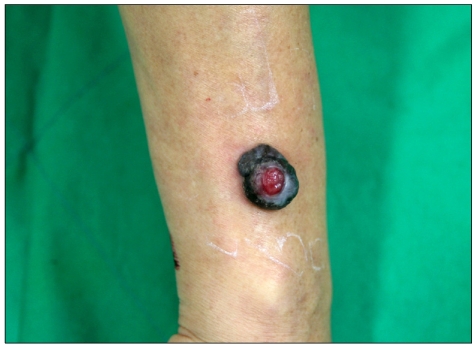
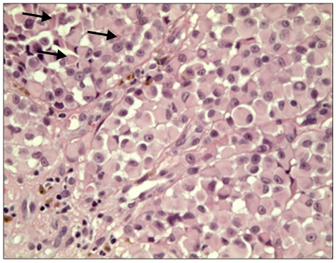
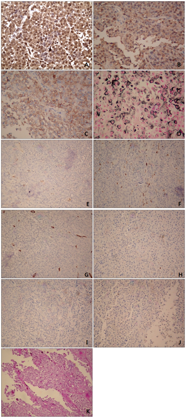
Melanoma with rhabdoid features is an uncommon variant of malignant melanoma. Here, we describe a rare case of primary rhabdoid malignant melanoma. A 54-year-old man presented with a black tumor measuring 3×4 cm on the right forearm. Histologic sections showed a tumor mass with rhabdoid features composed entirely of polygonal neoplastic cells with eccentric nuclei, prominent nucleoli, and large hyaline cytoplasmic inclusions. The tumor cells were immunoreactive with HMB-45, S100, Fontana-Masson silver and vimentin, and negative for smooth muscle actin, CD68, CD34, CD99, synaptophysin, desmin, and PAS. The differential diagnosis for this tumor included malignant peripheral nerve sheath tumor, malignant peripheral neuroectodermal tumor and rhabdomyosarcoma. The patient was treated with a wide excision and a local skin graft. The excised tumor was entirely composed of rhabdoid tumor cells. No recurrence or metastasis was evident 4 months after removal. This article is relevant to rare cases of primary malignant melanomas showing rhabdoid tumor cells over the entire excised lesion.
Keywords: Malignant melanoma; Rhabdoid.
Figures

References
-
- Haas JE, Palmer NF, Weinberg AG, Beckwith JB. Ultrastructure of malignant rhabdoid tumor of the kidney. A distinctive renal tumor of children. Hum Pathol. 1981;12:646–657. - PubMed
-
- Schmidt D, Harms D, Zieger G. Malignant rhabdoid tumor of the kidney. Histopathology, ultrastructure and comments on differential diagnosis. Virchows Arch A Pathol Anat Histopathol. 1982;398:101–108. - PubMed
-
- Gavino AC, Gillies EM. Metastatic rhabdoid melanoma: report of a case with a comparative review of the literature. J Cutan Pathol. 2008;35:337–342. - PubMed
-
- Bittesini L, Dei Tos AP, Fletcher CD. Metastatic malignant melanoma showing a rhabdoid phenotype: further evidence of a non-specific histological pattern. Histopathology. 1992;20:167–170. - PubMed
-
- Magro CM, Crowson AN, Mihm MC. Unusual variants of malignant melanoma. Mod Pathol. 2006;19(Suppl 2):S41–S70. - PubMed
Publication types
LinkOut - more resources
Full Text Sources
