

Cutaneous metaplastic synovial cyst of the first metatarsal head area
- PMID: 22148041
- PMCID: PMC3229056
- DOI: 10.5021/ad.2011.23.S2.S165
Cutaneous metaplastic synovial cyst of the first metatarsal head area
Abstract
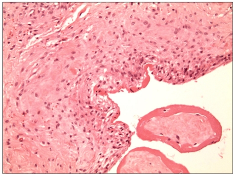
A cutaneous metaplastic synovial cyst (CMSC) is a cyst lined with metaplastic synovial tissue, which includes the formation of an intracystic villous structure resembling hyperplastic synovial villi. Clinically, the lesion is a tender, subcutaneous nodule that usually occurs at the site of previous surgical trauma and is frequently misdiagnosed as a suture granuloma. The actual cause remains unclear; however, trauma is presumed to be a precipitating factor, as most reported cases have demonstrated a history of antecedent cutaneous injury. Here, we present a case of CMSC in a 51-year-old woman who presented with a cystic mass localized in the left sole. She had no history of previous trauma or surgical procedures performed in the area. Although the case explained in this report is a spontaneous case of CMSC that occurred without a history of trauma, it is believed to have been caused by constant and chronic pressure since CMSC occurred in the first metatarsal head area, a part of the sole where heavy pressure is consistently applied.
Keywords: Chronic pressure; Cutaneous metaplastic synovial cyst; Sole.
Figures


References
-
- Gonzalez JG, Chiselli RW, Santa Cruz DJ. Synovial metaplasia of the skin. Am J Surg Pathol. 1987;11:343–350. - PubMed
-
- Bhawan J, Dayal Y, González-Serva A, Eisen R. Cutaneous metaplastic synovial cyst. J Cutan Pathol. 1990;17:22–26. - PubMed
-
- Nieto S, Buezo GF, Jones-Caballero M, Fraga J. Cutaneous metaplastic synovial cyst in an Ehlers-Danlos patient. Am J Dermatopathol. 1997;19:407–410. - PubMed
-
- Stern DR, Sexton FM. Metaplastic synovial cyst after partial excision of nevus sebaceous. Am J Dermatopathol. 1988;10:531–535. - PubMed
-
- Gómez Dorronsoro ML, Martinez-Peñuela JM, Ruiz de la Hermosa J. Metaplastic synovial cyst. Am J Surg Pathol. 1988;12:649–650. - PubMed
Publication types
LinkOut - more resources
Full Text Sources
