

Late-onset eccrine angiomatous hamartoma associated with a ganglion cyst on the sole of the foot
- PMID: 22148055
- PMCID: PMC3229070
- DOI: 10.5021/ad.2011.23.S2.S218
Late-onset eccrine angiomatous hamartoma associated with a ganglion cyst on the sole of the foot
Abstract
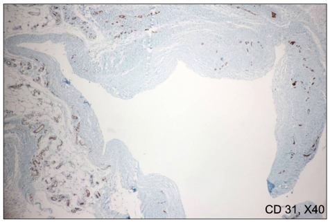
Eccrine angiomatous hamartoma (EAH) is a benign, uncommon, combined vascular and eccrine malformation. Most cases of this disorder have been single or multiple nodules or plaques that appear red, yellow, blue, violaceous, or skin colored. EAH may be congenital or appear later in childhood; it rarely arises during puberty or adulthood. A 52-year-old female patient visited our department for tender subcutaneous cystic tumor on the right sole with a one month history. Histopathologic examination confirmed EAH. During excisional biopsy procedure, mucinous discharges were observed which were histopathologically diagnosed as ganglion.
Keywords: Eccrine angiomatous hamartoma; Ganglion; Late-onset; Sole.
Figures


Similar articles
-
Eccrine angiomatous hamartoma: a review of ten cases.Ann Dermatol. 2013 May;25(2):208-12. doi: 10.5021/ad.2013.25.2.208. Epub 2013 May 10. Ann Dermatol. 2013. PMID: 23717013 Free PMC article.
-
Adult-Onset Subungual Eccrine Angiomatous Hamartoma on Right Great Toe: A Case Report.Ann Dermatol. 2023 Nov;35(Suppl 2):S265-S267. doi: 10.5021/ad.22.011. Ann Dermatol. 2023. PMID: 38061718 Free PMC article.
-
Multiple mucinous and lipomatous variant of eccrine angiomatous hamartoma associated with spindle cell hemangioma: a novel collision tumor?J Cutan Pathol. 2006 Apr;33(4):323-6. doi: 10.1111/j.0303-6987.2006.00413.x. J Cutan Pathol. 2006. PMID: 16630186
-
Congenital eccrine angiomatous hamartoma: Expanding the morphologic presentation and a review of the literature.Pediatr Dermatol. 2019 Nov;36(6):909-912. doi: 10.1111/pde.13974. Epub 2019 Aug 13. Pediatr Dermatol. 2019. PMID: 31410905 Review.
-
Eccrine angiomatous hamartoma: a report of symmetric and painful lesions of the wrists.Pediatr Dermatol. 2001 Mar-Apr;18(2):117-9. doi: 10.1046/j.1525-1470.2001.018002117.x. Pediatr Dermatol. 2001. PMID: 11358550 Review.
Cited by
-
Eccrine angiomatous hamartoma: a review of ten cases.Ann Dermatol. 2013 May;25(2):208-12. doi: 10.5021/ad.2013.25.2.208. Epub 2013 May 10. Ann Dermatol. 2013. PMID: 23717013 Free PMC article.
References
-
- Lotzbeck C. Ein Fall von Schweissdrsengeschwulst an der Wauge. Virchow Arch Pathol Anat Physiol Klin Med. 1859;16:160.
-
- Hyman AB, Harris H, Brownstein MH. Eccrine angiomatous hamartoma. N Y State J Med. 1968;68:2803–2806. - PubMed
-
- Pelle MT, Pride HB, Tyler WB. Eccrine angiomatous hamartoma. J Am Acad Dermatol. 2002;47:429–435. - PubMed
-
- Martinelli PT, Tschen JA. Eccrine angiomatous hamartoma: a case report and review of the literature. Cutis. 2003;71:449–455. - PubMed
-
- Zeller DJ, Goldman RL. Eccrine-pilar angiomatous hamartoma. Report of a unique case. Dermatologica. 1971;143:100–104. - PubMed
Publication types
LinkOut - more resources
Full Text Sources
