doi: 10.5056/jnm.2011.17.4.416.
Epub 2011 Oct 31.
How to interpret a functional or motility test - defecography
Affiliations
- PMID: 22148112
- PMCID: PMC3228983
- DOI: 10.5056/jnm.2011.17.4.416
Item in Clipboard
How to interpret a functional or motility test - defecography
J Neurogastroenterol Motil.
2011 Oct.
Abstract
Defecography evaluates in real time the morphology of rectum and anal canal in a physiologic setting by injection of a thick barium paste into the rectum and its subsequent evacuation. Because of its ability of structural and functional evaluation, defecography is primarily performed for work up of patients with longstanding constipation, unexplained anal or rectal pain, residual sensation after defecation or suspected prolapse. Technique and interpretation of this examination are outlined in this review.
Keywords: Constipation; Defecation; Defecography; Pelvic floor.
Conflict of interest statement
Conflicts of interest: None.
Figures
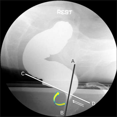
Measurement of anorectal angle. Anorectal angle (curved arrow) is measured between the longitudinal axis of anal canal (AB) and the posterior rectum line parallel to the rectum longitudinal axis (CD). Double thin arrows show the position of the anorectal junction.

Normal defecography. At rest (A). Note the deeper impression exerted by the puborectal sling (arrow) and the cranial migration of the distal rectum during forced contraction (B). During straining with closed sphincters (C), caudal migration of the anorectal junction is seen (asterisk). During evacuation (D), the anal canal opens with loss of puborectalis impression.

Dyskinetic puborectalis muscle syndrome. Note abnormally deep puborectal impression (arrow) at rest (A) and at evacuation phase (B). During evacuation phase, there is lack of pelvic floor descent.
Similar articles
-
MR Defecography in Assessing Functional Defecation Disorder: Diagnostic Value of the Defecation Phase in Detection of Dyssynergic Defecation and Pelvic Floor Prolapse in Females.Digestion. 2019;100(2):109-116. doi: 10.1159/000494249. Epub 2019 Jan 29. Digestion. 2019. PMID: 30695788
-
[Scintigraphic assessment of the anorectal function in children with idiopathic constipation].Zhonghua Er Ke Za Zhi. 2004 May;42(5):358-61. Zhonghua Er Ke Za Zhi. 2004. PMID: 15189695 Chinese.
-
Anorectal function in patients with defecation disorders and asymptomatic subjects: evaluation with defecography.Radiology. 1990 Jan;174(1):121-3. doi: 10.1148/radiology.174.1.2294537. Radiology. 1990. PMID: 2294537
-
[Anorectal functional study. The state of the art].Minerva Chir. 1994 Dec;49(12):1187-93. Minerva Chir. 1994. PMID: 7746437 Review. Italian.
-
Evacuation proctography (defecography): an aid to the investigation of pelvic floor disorders.Obstet Gynecol. 1994 Feb;83(2):307-14. Obstet Gynecol. 1994. PMID: 8290201 Review.
Cited by
-
Frequency, spectrum, and factors associated with fecal evacuation disorders among patients with chronic constipation referred to a tertiary care center in northern India.Indian J Gastroenterol. 2016 Mar;35(2):83-90. doi: 10.1007/s12664-016-0631-6. Epub 2016 Apr 4. Indian J Gastroenterol. 2016. PMID: 27041380
-
Chronic constipation in Rome IV era: The Indian perspective.Indian J Gastroenterol. 2017 May;36(3):163-173. doi: 10.1007/s12664-017-0757-1. Epub 2017 Jun 23. Indian J Gastroenterol. 2017. PMID: 28643273 Review.
-
Understanding and treating refractory constipation.World J Gastrointest Pharmacol Ther. 2014 May 6;5(2):77-85. doi: 10.4292/wjgpt.v5.i2.77. World J Gastrointest Pharmacol Ther. 2014. PMID: 24868488 Free PMC article. Review.
-
Fecal Evacuation Disorder Among Patients With Solitary Rectal Ulcer Syndrome: A Case-control Study.J Neurogastroenterol Motil. 2014 Oct 30;20(4):531-8. doi: 10.5056/jnm14030. J Neurogastroenterol Motil. 2014. PMID: 25273123 Free PMC article.
-
Anorectal Physiology Testing for Prolapse-What Tests are Necessary?Clin Colon Rectal Surg. 2021 Jan;34(1):15-21. doi: 10.1055/s-0040-1714246. Epub 2020 Sep 4. Clin Colon Rectal Surg. 2021. PMID: 33536845 Free PMC article. Review.
References
-
- Brodén B, Snellman B. Procidentia of the rectum studied with cineradiography. A contribution to the discussion of causative mechanism. Dis Colon Rectum. 1968;11:330–347. - PubMed
-
- Mahieu PH, Pringot J, Bodart P. Defecography: I. Description of a new procedure and results in normal patients. Gastrointest Radiol. 1984;9:247–251. - PubMed
-
- Mahieu PH, Pringot J, Bodart P. Defecograph: II. Contribution to the diagnosis of defection disorders. Gastrointest Radiol. 1984;9:253–261. - PubMed
-
- Ganeshan A, Anderson EM, Upponi S, et al. Imaging of obstructed defecation. Clin Radiol. 2008;63:18–26. - PubMed
-
- Roos JE, Weishaupt D, Wildermuth S, Willmann JK, Marincek B, Hilfiker PR. Experience of 4 years with open MR defecography: pictorial review of anorectal anatomy and disease. Radiographics. 2002;22:817–832. - PubMed
LinkOut - more resources
Full Text Sources