. 2012 Jan 11;134(1):173-5.
doi: 10.1021/ja210273w. Epub 2011 Dec 15.

Effects of DNA methylation on the structure of nucleosomes

Affiliations

Effects of DNA methylation on the structure of nucleosomes

Ju Yeon Lee et al. J Am Chem Soc. .

Abstract

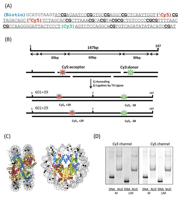
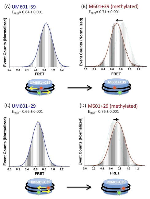
Nucleosomes are the fundamental packing units of the eukaryotic genome. Understanding the dynamic structure of a nucleosome is a key to the elucidation of genome packaging in eukaryotes, which is tied to the mechanisms of gene regulation. CpG methylation of DNA is an epigenetic modification associated with the inactivation of transcription and the formation of a repressive chromatin structure. Unraveling the changes in the structure of nucleosomes upon CpG methylation is an essential step toward the understanding of the mechanisms of gene repression and silencing by CpG methylation. Here we report single-molecule and ensemble fluorescence studies showing how the structure of a nucleosome is affected by CpG methylation. The results indicate that CpG methylation induces tighter wrapping of DNA around the histone core accompanied by a topology change. These findings suggest that changes in the physical properties of nucleosomes induced upon CpG methylation may contribute directly to the formation of a repressive chromatin structure.


Figures

Figure 1
Figure 1
Nucleosomal DNA constructs based on the Selex 601 sequence and nucleosome reconstitution. (A) Selex 601 DNA sequence. Locations of Cy3 (FRET donor) and Cy5 (FRET acceptor) are marked. 1Cy5 is at the +39th base from the dyad (Cytosine, 74th) (601+39 DNA) and 2Cy5 is at the +29th base from the dyad (601+29 DNA). Cy3 is located at the −38th base from the dyad. Underlined sequence represents the double-strand DNA region. (B) The 147 bp DNA fragment comprises ~60 bp of (H3-H4)2 tetramer binding site at the center and ~30 bp of H2A-H2B dimer binding sites near the termini. (C) The locations of fluorophores (Cy3 (Green), 1Cy5 (Red), 2Cy5 (Purple)) and methylation (black) in a crystal structure (PDB: 3MVD) is shown in a sphere representation. (D) Unmethylated (UM) or methylated (M) nucleosomes were reconstituted with yeast nucleosome assembly protein 1 (Nap1) and analyzed on a 5% native polyacrylamide gel.
FIGURE 2
FIGURE 2
Effects of CpG methylation on the structure of assembled nucleosomes. FRET efficiency histograms of (A) unmethylated 601+39 nucleosomes, (B) methylated 601+39 nucleosomes, (C) unmethylated 601+29 nucleosomes, and (D) methylated 601+29 nucleosomes. The pale-colored histograms in (B) and (D) are the replicas of the histograms in (A) and (C), respectively.

References

    1. Kornberg RD, Klug A. Sci Am. 1981;244:52. - PubMed
    1. Luger K, Mader AW, Richmond RK, Sargent DF, Richmond TJ. Nature. 1997;389:251. - PubMed
    1. Anselmi C, Bocchinfuso G, De Santis P, Savino M, Scipioni A. Biophys J. 2000;79:601. - PMC - PubMed
    1. Widom J. Quart Rev Biophys. 2001;34:269. - PubMed
    1. Segal E, Fondufe-Mittendorf Y, Chen L, Thastrom AC, Field Y, Moore IK, Wang JPZ, Widom J. Nature. 2006;442:772. - PMC - PubMed

Publication types