. 2012 Jan 10;51(1):329-41.
doi: 10.1021/bi201380p. Epub 2011 Dec 30.

Cyclostreptin derivatives specifically target cellular tubulin and further map the paclitaxel site

Affiliations

Cyclostreptin derivatives specifically target cellular tubulin and further map the paclitaxel site

Enrique Calvo et al. Biochemistry. .

Abstract

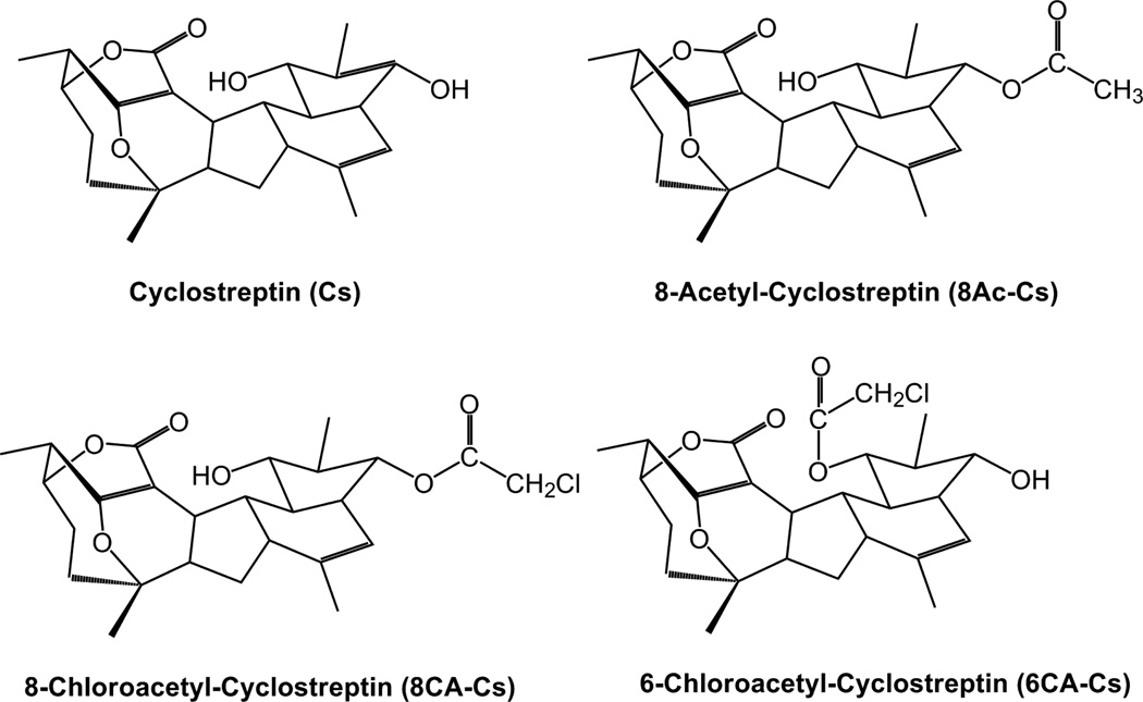
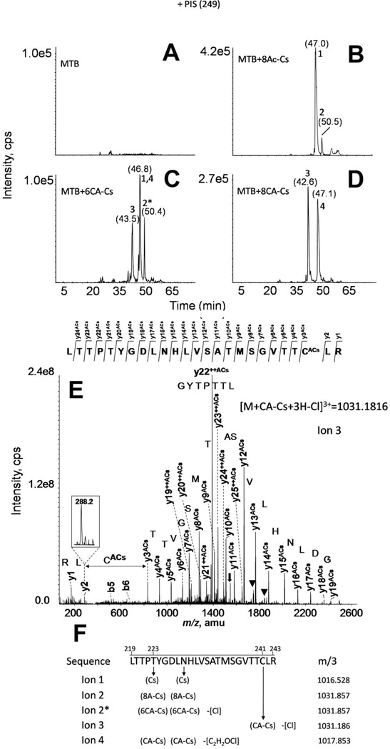
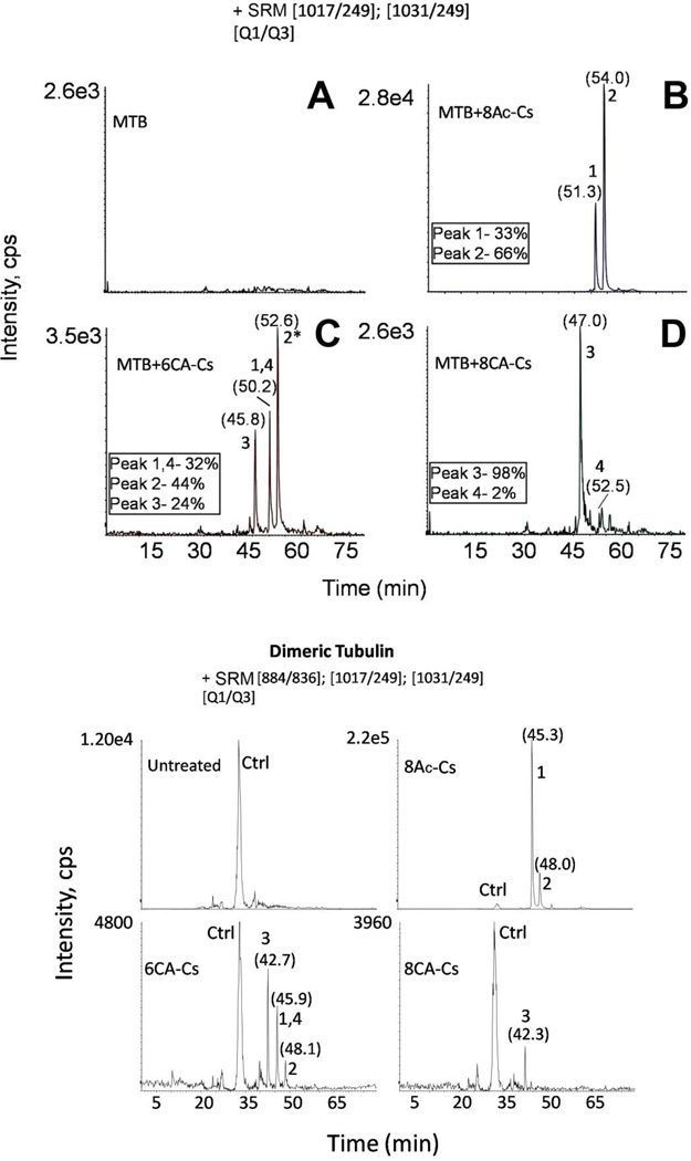
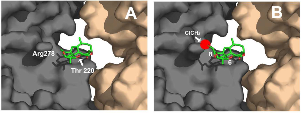
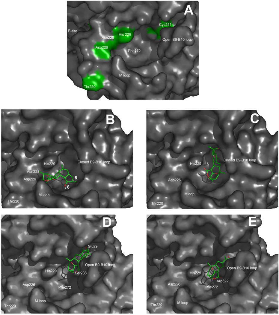
Cyclostreptin is the first microtubule-stabilizing agent whose mechanism of action was discovered to involve formation of a covalent bond with tubulin. Treatment of cells with cyclostreptin irreversibly stabilizes their microtubules because cyclostreptin forms a covalent bond to β-tubulin at either the T220 or the N228 residue, located at the microtubule pore or luminal taxoid binding site, respectively. Because of its unique mechanism of action, cyclostreptin overcomes P-glycoprotein-mediated multidrug resistance in tumor cells. We used a series of reactive cyclostreptin analogues, 6-chloroacetyl-cyclostreptin, 8-chloroacetyl-cyclostreptin, and [(14)C-acetyl]-8-acetyl-cyclostreptin, to characterize the cellular target of the compound and to map the binding site. The three analogues were cytotoxic and stabilized microtubules in both sensitive and multidrug resistant tumor cells. In both types of cells, we identified β-tubulin as the only or the predominantly labeled cellular protein, indicating that covalent binding to microtubules is sufficient to prevent drug efflux mediated by P-glycoprotein. 6-Chloroacetyl-cyclostreptin, 8-chloroacetyl-cyclostreptin, and 8-acetyl-cyclostreptin labeled both microtubules and unassembled tubulin at a single residue of the same tryptic peptide of β-tubulin as was labeled by cyclostreptin (219-LTTPTYGDLNHLVSATMSGVTTCLR-243), but labeling with the analogues occurred at different positions of the peptide. 8-Acetyl-cyclostreptin reacted with either T220 or N228, as did the natural product, while 8-chloroacetyl-cyclostreptin formed a cross-link to C241. Finally, 6-chloroacetyl-cyclostreptin reacted with any of the three residues, thus labeling the pathway for cyclostreptin-like compounds, leading from the pore where these compounds enter the microtubule to the luminal binding pocket.


Figures

Figure 1
Figure 1
Chemical structure of the Cs derivatives.
Figure 2
Figure 2
Upper Panel. Effect of Cs and derivatives on the cytoplasmic MTs of A549 cells. Cells were incubated either with drug solvent (DMSO) (control cells) (A), 500 nM Cs (B), 2.5 µM 8Ac-Cs (C), 2.5 µM [14C]8Ac-Cs (D), 5 µM 6CA-Cs (E) or 5 µM 8CA-Cs (F). MTs were immunostained with an α-tubulin monoclonal antibody, and DNA was stained with Hoechst 33342. Insets are mitotic cells from the same preparation. The scale bar represents 10 µm. Lower Panel. Effect of Cs and derivatives on the cell cycle of 1A9, A2780AD and A549 cells. Cells were incubated either with drug solvent (DMSO) (control cells) (A), 100 nM Cs (B), 400 nM 8Ac-Cs (C), 400 nM [14C]8Ac-Cs (D), 200 nM 6CA-Cs (E) or 400 nM 8CA-Cs (F). The concentrations are those where maximal accumulation of cells in G2/M phase was observed. PI, propidium iodide.
Figure 3
Figure 3
Upper Panel Identification of [14C]8Ac-Cs protein cell targets. Two dimensional gel electrophoresis of proteins extracted from A549 cells incubated with 2.5 µM [14C]8Ac-Cs for 24 h. (A) Autoradiogram of the PDVF membrane after protein transfer. (B) The silver-stained 2D replica gel obtained with the protein extracts from treated cells and the spots corresponding to radiolabeled proteins are indicated (1 to 4). Protein identification was performed by in-gel trypsin digestion followed by database search with MALDI-TOF-MS data. Panels C,D,E.-[14C]8Ac-Cs cell target is conserved in different cell lines. 1A9 cells incubated with 2.5 µM (C) or 300 nM [14C]8Ac-Cs (D) and A549 cells incubated with 300 nM [14C]8Ac-Cs (E). Lower Panel. HPLC analysis of 6CA-Cs extracted from pellets (red) and supernatants (black) after incubation without (F) and with (G) stabilized MTs.
Figure 4
Figure 4
MS analyses of Cs derivative binding to assembled MTs by PIS. Total ion chromatograms of the PIS experiments using the diagnostic ion at 249 m/z from control (A), or Cs derivative-treated, trypsin-digested MTs (MTB): 8Ac-Cs (B), 6CA-Cs (C), and 8CA-Cs (D). (E) Fragmentation spectrum from ion 3. Main fragmentation series (y-carboxy and b-amino) are indicated. Numbers above chromatographic peaks indicate the retention time (in brackets) and the type of detected ion. Water loss ions are indicated with black triangles. Doubly-charged fragment ions are labeled with ++, and a black arrow indicates the doubly-charged parent ion. (F) Hypothetical structure of Cs derivative adducts. A schematic structure for the hypothetical structure of ions 1 to 4 is presented. In brackets are marked the linked group in each case attached to the corresponding residue through an arrow. The group loss in the different linkages is labeled in square brackets. m/3 indicate the exact mass observed for the triply-charged tubulin-derived adducts.
Figure 5
Figure 5
Upper Panel Estimation of adduct abundance by SRM. Extracted ion chromatogram for the two ion pairs analyzed corresponding to Cs derivative linked adducts. The relative intensity percentage (numbered boxes) is indicated for ions detected in 8Ac-Cs-(B), 6CA-Cs- (C), and 8CA-Cs-treated MT samples (D). (A) Untreated MT sample. Numbers above chromatographic peaks indicate the retention time (in brackets) and the type of ion detected. Lower Panel MS analyses of Cs derivative binding to dimeric tubulin by SRM. Extracted ion chromatogram for SRM experiments of 3 ion pairs, including the ion pair corresponding to the tubulin-derived unmodified tryptic peptide (Q1 884 m/z, Q3 836 m/z, labeled as Ctrl) in dimeric tubulin samples. Numbers above chromatographic peaks indicate the retention time (in brackets) and the type of detected ion.
Figure 6
Figure 6
Molecular models of: (A) Cs bound to Thr220 in the pore binding site of MTs as described in (20) and (B) 8CA-Cs placed in the same binding pose to show the steric hindrance that would result from the presence of the chloroacetyl moiety at position 8 (red sphere).
Figure 7
Figure 7
Molecular models of: (A) The extended luminal pore site of PTX in MTs, (B) Cs docked into the luminal site (first pose), (C) Cs docked into the luminal site (second pose). (D) 6CA-Cs bound to Cys241 in the extended luminal site and (E) 8CA-Cs bound to Cys241 in the extended luminal site.

References

    1. Jemal A, Thomas A, Murray T, Thun M. Cancer statistics, 2002. CA Cancer J Clin. 2002;52:23–47. - PubMed
    1. Schiff PB, Fant J, Horwitz SB. Promotion of microtubule assembly in vitro by taxol. Nature. 1979;277:665–667. - PubMed
    1. Galsky MD, Dritselis A, Kirkpatrick P, Oh WK. Cabazitaxel. Nat Rev Drug Discov. 2010;9:677–678. - PubMed
    1. Dumontet C, Jordan MA, Lee FF. Ixabepilone: targeting βIII-tubulin expression in taxane-resistant malignancies. Mol Cancer Ther. 2009;8:17–25. - PubMed
    1. Pryor DE, O'Brate A, Bilcer G, Díaz JF, Wang Y, Kabaki M, Jung MK, Andreu JM, Ghosh AK, Giannakakou P, Hamel E. The microtubule stabilizing agent laulimalide does not bind in the taxoid site, kills cells resistant to paclitaxel and epothilones, and may not require its epoxide moiety for activity. Biochemistry. 2002;41:9109–9115. - PubMed

Publication types

MeSH terms