. 2011 Dec;38(12):6754-62.
doi: 10.1118/1.3663675.

Commissioning a small-field biological irradiator using point, 2D, and 3D dosimetry techniques

Affiliations

Commissioning a small-field biological irradiator using point, 2D, and 3D dosimetry techniques

Joseph Newton et al. Med Phys. 2011 Dec.

Abstract

Purpose: To commission a small-field biological irradiator, the XRad225Cx from Precision x-Ray, Inc., for research use. The system produces a 225 kVp x-ray beam and is equipped with collimating cones that produce both square and circular radiation fields ranging in size from 1 to 40 mm. This work incorporates point, 2D, and 3D measurements to determine output factors (OF), percent-depth-dose (PDD) and dose profiles at multiple depths.

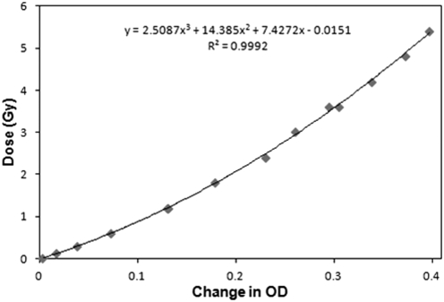
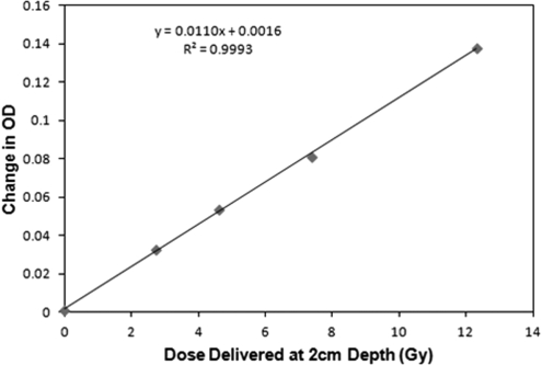
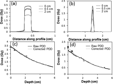
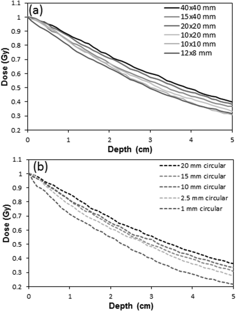
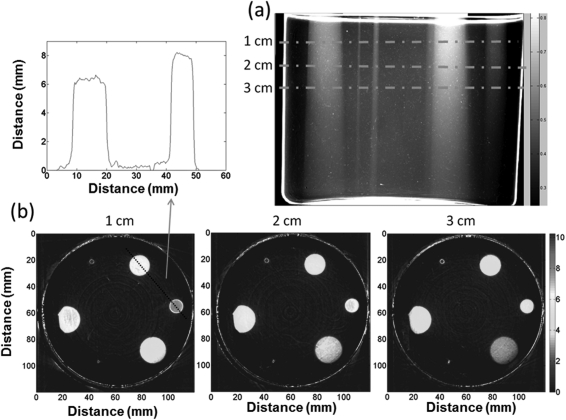
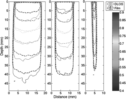
Methods: Three independent dosimetry systems were used: ion-chambers (a farmer chamber and a micro-ionisation chamber), 2D EBT2 radiochromic film, and a novel 3D dosimetry system (DLOS∕PRESAGE®). Reference point dose rates and output factors were determined from in-air ionization chamber measurements for fields down to ∼13 mm using the formalism of TG61. PDD, profiles, and output factors at three separate depths (0, 0.5, and 2 cm), were determined for all field sizes from EBT2 film measurements in solid water. Several film PDD curves required a scaling correction, reflecting the challenge of accurate film alignment in very small fields. PDDs, profiles, and output factors were also determined with the 3D DLOS∕PRESAGE® system which generated isotropic 0.2 mm data, in scan times of 20 min.

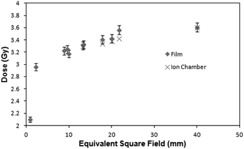
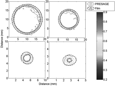
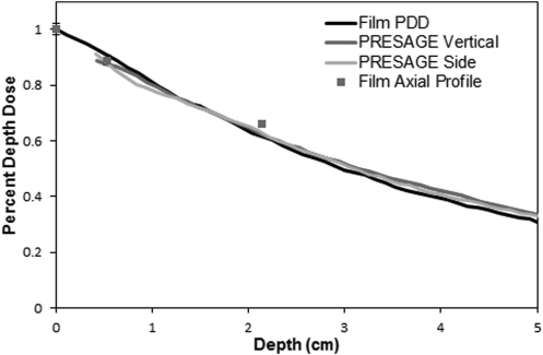
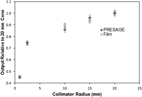
Results: Surface output factors determined by ion-chamber were observed to gradually drop by ∼9% when the field size was reduced from 40 to 13 mm. More dramatic drops were observed for the smallest fields as determined by EBT∼18% and ∼42% for the 2.5 mm and 1 mm fields, respectively. PRESAGE® and film output factors agreed well for fields <20 mm (where 3D data were available) with mean deviation of 2.2% (range 1%-4%). PDD values at 2 cm depth varied from ∼72% for the 40 mm field, down to ∼55% for the 1 mm field. EBT and PRESAGE® PDDs agreed within ∼3% in the typical therapy region (1-4 cm). At deeper depths the EBT curves were slightly steeper (2.5% at 5 cm). These results indicate good overall consistency between ion-chamber, EBT2 and PRESAGE® measured OFs, PDDs, and profiles.

Conclusions: The combination of independent 2D and 3D measurements was found to be valuable to ensure accurate and comprehensive commissioning. Film measurements were time consuming and challenging due to the difficulty of film alignment in small fields. PRESAGE® 3D measurements were comprehensive and efficient, because alignment errors are negligible, and all parameters for multiple fields could be obtained from a single dosimeter and scan. However, achieving accurate superficial data (within 4 mm) is not yet feasible due to optical surface artifacts.


Figures

Figure 1
Figure 1
EBT2 calibration curve measured with the 40 × 40 mm2 field. Optical densities (OD) are converted to dose using the fit equation.
Figure 2
Figure 2
PRESAGE® calibration curve determined from four irradiations of the calibration dosimeter, with the 15 mm diameter field.
Figure 3
Figure 3
Absolute output factors (60 s irradiation with a 225 kVp 13 mA beam) determined at isocenter at the water surface (SSD = 30.76 cm).
Figure 4
Figure 4
Beam profiles for the 15 mm circular field (a) and 1 mm circular field (b) at depths of 0 cm, 0.5 cm and 2 cm in solid water. (c) The PDD curve for the 15 mm circular field, and (d) for the 1 mm field. The three data markers in c and d indicate data from axial films where alignment errors were negligible.
Figure 5
Figure 5
Corrected EBT2 PDD curves for the six rectangular fields (a) and five circular fields (b) obtained with film aligned to the central axis and sandwiched between solid water blocks.
Figure 6
Figure 6
(a) An illustrative projection image of the PRESAGE® dosimeter irradiated with five circular fields (diameter 20, 15, 10, 2.5, and 1mm) incident on the top surface. The full reconstructed dose cube was 0.2 mm3 voxel size throughout the whole dosimeter. Three representative axial planes are shown in (b) along with a line profile through the center of the 15 and 10 mm fields.
Figure 7
Figure 7
Isodose contour plots of the 15 mm (upper left), 10 mm (upper right), 2.5 mm (lower left), and 1 mm (lower right) circular fields at a water-equivalent depth of 2 cm. Isodose lines are 90, 80, 50, and 20%.
Figure 8
Figure 8
PDD curves for the 15 mm circular field at 225 kVp measured in film, and PRESAGE (vertical end-on irradiation, and side irradiation). Each curve is corrected for effective depth in water. The values obtained from axial film measurements are shown as points.
Figure 9
Figure 9
Isodose contour plots (90, 80, 70, 60, 50, and 40%) of the PDD planes for the 15, 10, and 2.5 mm circular fields. The depths are density corrected for difference from water equivalency of both PRESAGE® and solid water.
Figure 10
Figure 10
PRESAGE® and film output factors relative to the 20 mm field film measurement measured at a depth of 2 cm.

References

    1. Kirsch D. G. et al., “p53 controls radiation-induced gastrointestinal syndrome in mice independent of apoptosis,” Science 327, 593–596 (2010).10.1126/science.1166202 - DOI - PMC - PubMed
    1. Kirsch D. G. et al., “Imaging primary lung cancers in mice to study radiation biology,” Int. J. Radiat. Oncol. Biol. Phys. 76, 973–977 (2010).10.1016/j.ijrobp.2009.11.038 - DOI - PMC - PubMed
    1. Thomas A. and Oldham M., “Fast, large field-of-view, telecentric optical-CT scanning system for 3D radiochromic dosimetry,” J. Phys. 250, 1–5 (2010).10.1088/1742-6596/250/1/0120 - DOI - PMC - PubMed
    1. Thomas A., Newton J. R., Oldham M., and Adamovics J., “Commissioning and benchmarking a 3D dosimetry system for clinical use,” Med. Phys. 38, 4846–4857 (2011).10.1118/1.3611042 - DOI - PMC - PubMed
    1. Das I. J. et al., “Accelerator beam data commissioning equipment and procedures: Report of the TG-106 of the Therapy Physics Committee of the AAPM,” Med. Phys. 35, 4186–4215 (2008).10.1118/1.2969070 - DOI - PubMed

Publication types