A case of semantic variant primary progressive aphasia with severe insular atrophy
- PMID: 22150361
- PMCID: PMC3500405
- DOI: 10.1080/13554794.2011.627343
A case of semantic variant primary progressive aphasia with severe insular atrophy
Abstract
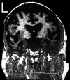
Insular degeneration has been linked to symptoms of frontotemporal dementia (FTD). Presented in this case is a patient exhibiting semantic variant primary progressive aphasia, behavioral disturbance. Upon autopsy, he was found to have severe insular atrophy. In addition, selective serotonin reuptake inhibitors were ineffective in reducing symptoms of obsessive-compulsive behaviors or emotional blunting. This case suggests that Seeley et al.'s (2007 , Alzheimer Disease & Associated Disorders, 21, S50) hypothesis that von Economo neurons and fork cell-rich brain regions, particularly in the insula, are targeted in additional subtypes of FTD beyond the behavioral variant.
Figures


References
-
- Penfield W. Functional localization in temporal and deep Sylvian areas. Res Publ Assoc Ner Ment Dis. 1956;36:210–226. - PubMed
-
- Gorno-Tempini ML, Pradelli S, Serafini M, et al. Explicit and incidental facial expression processing: an fMRI study. Neuroimage. 2001;14:465–473. - PubMed
-
- Woolley JD, Gorno-Tempini ML, Seeley WW, et al. Binge eating is associated with right orbitofrontal-insular-striatal atrophy in frontotemporal dementia. Neurology. 2007;69:1424–1433. - PubMed
Publication types
MeSH terms
Grants and funding
LinkOut - more resources
Full Text Sources
Miscellaneous
