

Callosal thickness reductions relate to facial dysmorphology in fetal alcohol spectrum disorders
- PMID: 22150665
- PMCID: PMC3309126
- DOI: 10.1111/j.1530-0277.2011.01679.x
Callosal thickness reductions relate to facial dysmorphology in fetal alcohol spectrum disorders
Abstract
Background: Structural abnormalities of the corpus callosum (CC), such as reduced size and increased shape variability, have been documented in individuals with fetal alcohol spectrum disorders (FASD). However, the regional specificity of altered CC structure, which may point to the timing of neurodevelopmental disturbances and/or relate to specific functional impairments, remains unclear. Furthermore, associations between facial dysmorphology and callosal structure remain undetermined.
Methods: One hundred and fifty-three participants (age range 8 to 16) including 82 subjects with FASD and 71 nonexposed controls were included in this study. The structural magnetic resonance imaging data of these subjects was collected at 3 sites (Los Angeles and San Diego, California, and Cape Town, South Africa) and analyzed using classical parcellation schemes, as well as more refined surface-based geometrical modeling methods, to identify callosal morphological alterations in FASD at high spatial resolution.
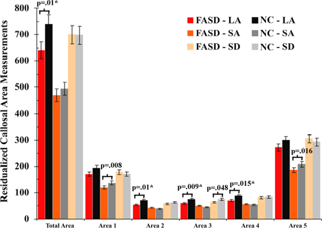
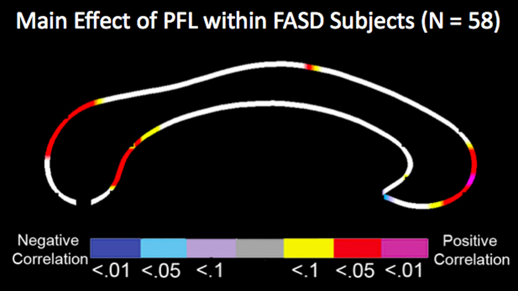
Results: Reductions in callosal thickness and area, specifically in the anterior third and the splenium, were observed in FASD compared with nonexposed controls. In addition, reduced CC thickness and area significantly correlated with reduced palpebral fissure length.
Conclusions: Consistent with previous reports, findings suggest an adverse effect of prenatal alcohol exposure on callosal growth and further indicate that fiber pathways connecting frontal and parieto-occipital regions in each hemisphere may be particularly affected. Significant associations between callosal and facial dysmorphology provide evidence for a concurrent insult to midline facial and brain structural development in FASD.
Copyright © 2011 by the Research Society on Alcoholism.
Conflict of interest statement
Figures

References
-
- Anderson MJ, Ter Braak CJF. Permutation tests for multi-factorial analysis of variance. Journal of Statistical Computation and Simulation. 2003;73:85–113.
-
- Archibald SL, Fennema-Notestine C, Gamst A, Riley EP, Mattson SN, Jernigan TL. Brain dysmorphology in individuals with severe prenatal alcohol exposure. Developmental Medicine and Child Neurology. 2001;43:148–154. - PubMed
-
- Astley SJ, Aylward EH, Olson HC, Kerns K, Brooks A, Coggins TE, Davies J, Dorn S, Gendler B, Jirikowic T, Kraegel P, Maravilla K, Richards T. Magnetic resonance imaging outcomes from a comprehensive magnetic resonance study of children with fetal alcohol spectrum disorders. Alcohol Clin Exp Res. 2009;33:1671–1689. - PMC - PubMed
-
- Astley SJ, Clarren SK. Diagnosing the full spectrum of fetal alcohol-exposed individuals: Introducing the 4-Digit Diagnostic Code. Alcohol and Alcoholism. 2000;35:400–410. - PubMed
-
- Bookstein FL, Sampson PD, Streissguth AP, Connor PD. Geometric morphometrics of corpus callosum and subcortical structures in the fetal-alcohol-affected brain. Teratology. 2001;64:4–32. - PubMed
Publication types
MeSH terms
Grants and funding
- U01 AA 11685/AA/NIAAA NIH HHS/United States
- U01 AA011685/AA/NIAAA NIH HHS/United States
- R01 AA015134/AA/NIAAA NIH HHS/United States
- U24 AA014811/AA/NIAAA NIH HHS/United States
- R01 DA017830/DA/NIDA NIH HHS/United States
- U01 AA017122/AA/NIAAA NIH HHS/United States
- R01DA017831/DA/NIDA NIH HHS/United States
- U24AA014811/AA/NIAAA NIH HHS/United States
- R01 HD053893/HD/NICHD NIH HHS/United States
- U01 AA014834/AA/NIAAA NIH HHS/United States
- U24 AA014808/AA/NIAAA NIH HHS/United States
- K99 MH093388/MH/NIMH NIH HHS/United States
- R01 HD053893-01/HD/NICHD NIH HHS/United States
- R01 AA15134/AA/NIAAA NIH HHS/United States
- U01 AA017122-01/AA/NIAAA NIH HHS/United States
LinkOut - more resources
Full Text Sources
Medical
Molecular Biology Databases
