Review
. 2012 May;36(3):684-705.
doi: 10.1111/j.1574-6976.2011.00320.x. Epub 2012 Jan 10.

The molecular basis of herpes simplex virus latency

Affiliations
Free PMC article
Review

The molecular basis of herpes simplex virus latency

Michael P Nicoll et al. FEMS Microbiol Rev. 2012 May.
Free PMC article

Abstract

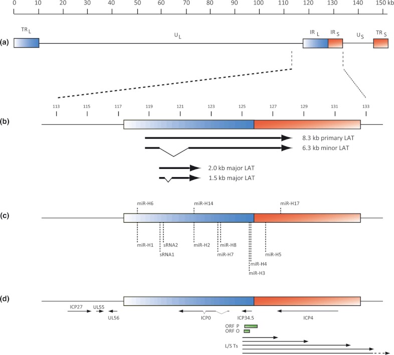
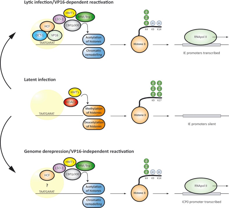
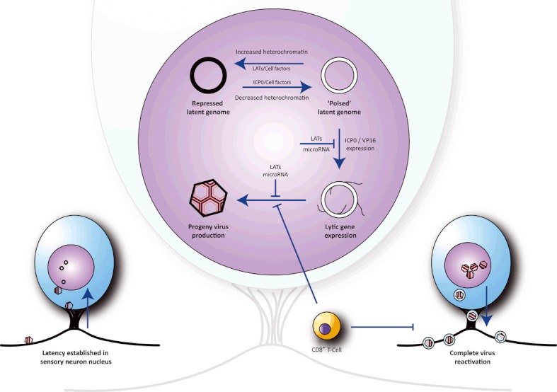
Herpes simplex virus type 1 is a neurotropic herpesvirus that establishes latency within sensory neurones. Following primary infection, the virus replicates productively within mucosal epithelial cells and enters sensory neurones via nerve termini. The virus is then transported to neuronal cell bodies where latency can be established. Periodically, the virus can reactivate to resume its normal lytic cycle gene expression programme and result in the generation of new virus progeny that are transported axonally back to the periphery. The ability to establish lifelong latency within the host and to periodically reactivate to facilitate dissemination is central to the survival strategy of this virus. Although incompletely understood, this review will focus on the mechanisms involved in the regulation of latency that centre on the functions of the virus-encoded latency-associated transcripts (LATs), epigenetic regulation of the latent virus genome and the molecular events that precipitate reactivation.


Figures

Fig 1
Fig 1
The genomic region of the HSV-1 LATs. (a) The prototypic organization of the HSV-1 genome. The 152 kb genome consists of the unique long (UL) and unique short (US) sequences, flanked by inverted repeat sequences termed the terminal and internal long and short repeats (TRL, IRL, TRS and IRS). (b) Enlarged view of the internal repeats displaying the coding location of the LAT RNA species. All LATs are processed from the 8.3 kb primary transcript, which is transcribed from a single promoter within the IRL. Above size markers represent kb. (c) Small noncoding RNAs expressed from within the LATs. With the exception of miR-H1, H6, H14 and H17, all displayed microRNA and sRNA are likely to be processed from the primary LAT transcript. (d) Lytic cycle virus genes encoded adjacent to or overlapping the LAT region. The L/ST transcripts represent a family of transcripts that are expressed late during productive infection from HSV-1 mutants lacking functional ICP4 (Yeh & Schaffer, 1993). Note that all genes displayed within the repeat sequences in b–d are diploid.
Fig 2
Fig 2
Chromatin control of HSV-1 gene expression. Two models of reactivation are shown, involving the specific activation of the virus regulatory proteins VP16 or ICP0 by cellular factors. Lytic infection/VP16-dependent reactivation: The VP16/Oct-1/HCF trimeric complex interacts with a number of co-activators including histone acetyltransferases (CBP/p300), chromatin remodelling factors (BGRF-1 and BRM), histone methyltransferases (HMT's) and lysine-specific demethylase-1 (LSD-1). Recognition of IE promoters by the VP16-induced complex results in activation of IE gene expression and prevention of repressive histone accumulation on the virus genome. Histone modifications associated with active transcription, such as H3K4me3 and H3K9ac, are associated with the virus genome during lytic infection. Latent infection: In the absence of virally encoded transactivators, HMT's and histone deacetylases (HDACs) maintain viral lytic promoters in a repressed chromatin state characterized with the accumulation of histone modifications associated with repression, such as H3K9me3, resulting in silencing of gene expression. An exception to this global silencing is the LAT locus, which is actively transcribed during latency. Genome depression/VP16-independent reactivation: In the absence of VP16, the presence of some or all cellular co-activators involved in the activation of IE promoters during lytic infection could facilitate gene expression following reversal of heterochromatin-based silencing. In this model, specific acetylation of the ICP0 promoter region would result in ICP0 gene expression, leading to further depression across the virus genome and reactivation.
Fig 3
Fig 3
The balance between latency and productive infection/reactivation is policed by cellular and viral factors. HSV latency is established in sensory neurons innervating the sit of primary infection. During the establishment of latency, within the cell nucleus, circularized viral DNA becomes associated with heterochromatin resulting in the repression of lytic cycle gene expression. Periodically, virus reactivation and exit from latency can occur as a consequence of changes in neuronal cell physiology in response to stress. Reversal of repressive histone modifications allows transcriptional activation of one or more viral gene products responsible for initiating virus replication. Accumulation of the LATs and cognate microRNAs during latency may facilitate establishment of latency and regulate reactivation by blocking translation of the viral IE genes ICP0 and ICP4. Ganglion-resident CD8+ T cells may actively halt reactivation through noncytolytic control mechanisms. Occasionally, reactivating cells escape cellular, virus and immune mechanisms of control leading to the completion of the virus life cycle, and production of progeny virus that is then transported to the periphery resulting in either asymptomatic or symptomatic infection that can lead to transmission of reactivated virus to immunologically naïve hosts.

References

    1. Ahmed M, Lock M, Miller CG, Fraser NW. Regions of the herpes simplex virus type 1 latency-associated transcript that protect cells from apoptosis in vitro and protect neuronal cells in vivo. J Virol. 2002;76:717–729. - PMC - PubMed
    1. Amelio AL, Giordani NV, Kubat NJ, O'neil JE, Bloom DC. Deacetylation of the herpes simplex virus type 1 latency-associated transcript (LAT) enhancer and a decrease in LAT abundance precede an increase in ICP0 transcriptional permissiveness at early times postexplant. J Virol. 2006;80:2063–2068. - PMC - PubMed
    1. Arthur J, Efstathiou S, Simmons A. Intranuclear foci containing low abundance herpes simplex virus latency-associated transcripts visualized by non-isotopic in situ hybridization. J Gen Virol. 1993;74(Pt 7):1363–1370. - PubMed
    1. Arthur JL, Scarpini CG, Connor V, Lachmann RH, Tolkovsky AM, Efstathiou S. Herpes simplex virus type 1 promoter activity during latency establishment, maintenance, and reactivation in primary dorsal root neurons in vitro. J Virol. 2001;75:3885–3895. - PMC - PubMed
    1. Bertke AS, Swanson SM, Chen J, Imai Y, Kinchington PR, Margolis TP. A5-positive primary sensory neurons are nonpermissive for productive infection with herpes simplex virus 1 in vitro. J Virol. 2011;85:6669–6677. - PMC - PubMed

Publication types