

Intrauterine growth and postnatal skeletal development: findings from the Southampton Women's Survey
- PMID: 22150706
- PMCID: PMC3641484
- DOI: 10.1111/j.1365-3016.2011.01237.x
Intrauterine growth and postnatal skeletal development: findings from the Southampton Women's Survey
Abstract
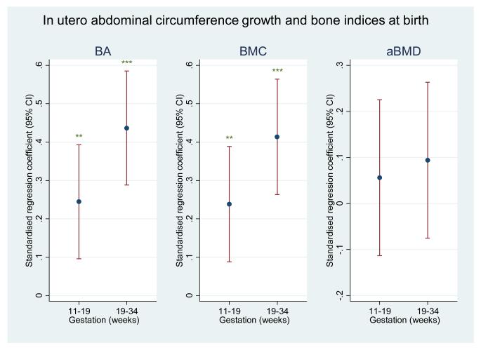
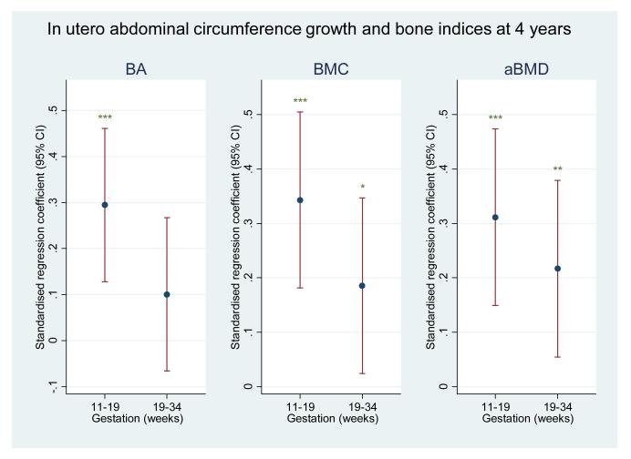
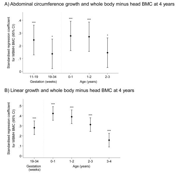
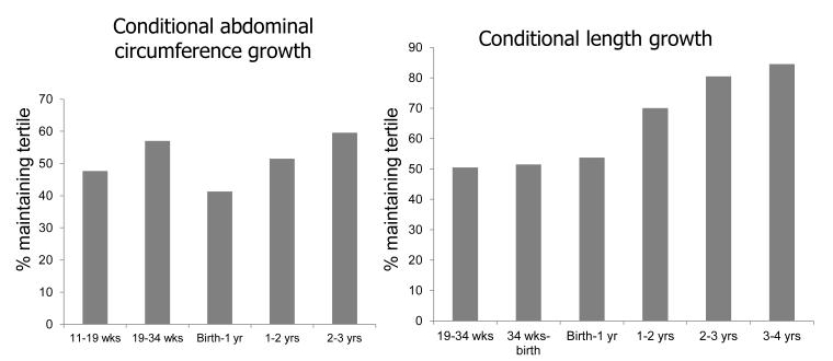
We have previously demonstrated associations between fetal growth in late pregnancy and postnatal bone mass. However, the relationships between the intrauterine and early postnatal skeletal growth trajectory remain unknown. We addressed this in a large population-based mother-offspring cohort study. A total of 628 mother-offspring pairs were recruited from the Southampton Women's Survey. Fetal abdominal circumference was measured at 11, 19 and 34 weeks gestation using high-resolution ultrasound with femur length assessed at 19 and 34 weeks. Bone mineral content was measured postnatally in the offspring using dual-energy X-ray absorptiometry at birth and 4 years; postnatal linear growth was assessed at birth, 6, 12, 24, 36 and 48 months. Late pregnancy abdominal circumference growth (19-34 weeks) was strongly (P < 0.01) related to bone mass at birth, but less robustly associated with bone mass at 4 years. Early pregnancy growth (11-19 weeks) was more strongly related to bone mass at 4 years than at birth. Postnatal relationships between growth and skeletal indices at 4 years were stronger for the first and second postnatal years, than the period aged 2-4 years. The proportion of children changing their place in the distribution of growth velocities progressively reduced with each year of postnatal life. The late intrauterine growth trajectory is a better predictor of skeletal growth and mineralisation at birth, while the early intrauterine growth trajectory is a more powerful determinant of skeletal status at age 4 years. The perturbations in this trajectory which influence childhood bone mass warrant further research.
© 2011 Blackwell Publishing Ltd.
Figures
References
-
- Tanner JM. Foetus into Man: Physical growth from conception to maturity. Castlemead Publications; Ware: 1989. Growth before birth; pp. 36–50.
-
- Tanner JM. Foetus into Man: Physical growth from conception to maturity. Castlemead Publications; Ware: 1989. The organisation of the growth process; pp. 165–177.
-
- Kovacs CS. Skeletal physiology: fetus and neonate. In: Favus MJ, editor. Primer on the metabolic bone diseases and disorders of mineral metabolism. ASBMR; Washington: 2003. pp. 65–71.
Publication types
MeSH terms
Grants and funding
LinkOut - more resources
Full Text Sources
Medical
