Review
. 2012 Jan;22(1):82-8.
doi: 10.1111/j.1750-3639.2011.00543.x.

Autophagy in lysosomal myopathies

Affiliations
Review

Autophagy in lysosomal myopathies

May Christine V Malicdan et al. Brain Pathol. 2012 Jan.

Abstract

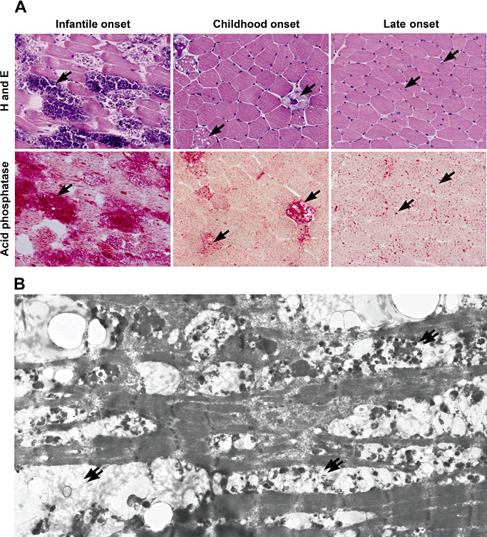
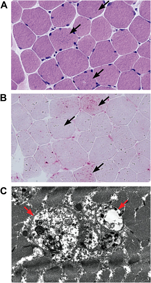
Lysosomal myopathies are hereditary myopathies characterized morphologically by the presence of autophagic vacuoles. In mammals, autophagy plays an important role for the turnover of cellular components, particularly in response to starvation or glucagons. In normal muscle, autolysosomes or autophagosomes are typically inconspicuous. In distinct neuromuscular disorders, however, lysosomes become structurally abnormal and functionally impaired, leading to the accumulation of autophagic vacuoles in myofibers. In some instances, the accumulation of autophagic vacuoles can be a prominent feature, implicating autophagy as a contributor to disease pathomechanism and/or progression. At present, there are two disorders in the muscle that are associated with a primary defect in lysosomal proteins, namely Pompe disease and Danon disease. This review will give a brief discussion on these disorders, highlighting the role of autophagy in disease progression.


Figures

Figure 1
Figure 1
Subtypes of Pompe diseases and findings in pathology. Hematoxylin and eosin staining (A) shows vacuolar structures (arrows) in myofibers pathognomonic for the disease especially in infantile onset form. In childhood onset form, several vacuoles are scattered all throughout the sections. The late‐onset form is characterized by milder changes, namely scattered inclusion bodies that may appear like rimmed vacuoles. The vacuolar structures are highly stained by acid phosphatase. Electron microscopy (B) shows that the vacuoles (double arrows) occupy the whole diameter of the fiber and disrupt myofibrillar structure.
Figure 2
Figure 2
Pathology changes in Danon disease. Hematoxylin and eosin (A) staining shows moderate variation in fiber size with small basophilic granules within the scattered myofibers (arrows). These granules are highlighted with acid phosphatase (B). In electron microscopy, the autophagic vacuoles with sarcolemmal features (AVSF) appear to be lined with a layer of basal lamina (red arrows) and contain small autophagic vacuoles, multilamellar bodies and electron dense material (C).
Figure 3
Figure 3
Histochemistry and immunohistochemistry. Transverse sections of skeletal muscle biopsies from Danon disease patients. Several fibers contain scattered tiny basophilic intracytoplasmic vacuoles (A): H&E. The vacuolar membrane has high nonspecific esterase (B) and acetylcholinesterase (C) activities. None of the vacuoles bind to α‐bungarotoxin (D). Sections were stained with antibody against the C‐terminus of dystrophin (E), the rod domain of dystrophin (F), the N‐terminus of dystrophin (G), laminin α2 (H), α‐sarcoglycan (I), β‐sarcoglycan (J), γ‐sarcoglycan (K), δ‐sarcoglycan (L), dystrobrevin (M), α‐dystroglycan (N), utrophin (O), dysferlin (P), β‐dystroglycan (Q), perlecan (R), caveolin‐E (S), collagen IV (T), fibronectin (U), collagen VI (V), integrin β1 (W), and agrin (X). The vacuolar membranes were immuno‐positive with most of the primary antibodies, although reactivity of these proteins was variable. The results are summarized in Table 1. Transverse 5‐μm serial sections (Y1–Y5) and longitudinal section (Z) of muscle from Danon disease patient showing immunoreaction for dystrophin. Vacuolar membrane in muscle fiber (*) is not connected to the sarcolemma but is closed. Longitudinal section shows that the vacuoles are spherical or oval. (D–W, Y1–Y5, Z): FIT C‐labeled staining; (X): DAB staining, (C–S, U, V, Y1–Y5): serial sections. Scale bars: (A–W, Y1–Y5)= 20 μm; (Z)= 30 μm. Reproduced with permission from Kazuma S et al (2005) Autophagic vacuoles with sarcolemmal features delineate Danon disease and related myopathies. 64: 513–522.

References

    1. Akanas V, Engel WK (2006) Inclusion‐body myositis: a myodegenerative conformational disorder associated with Abeta, protein misfolding, and proteasome inhibition. Neurology 66:S39–S48. - PubMed
    1. Amalfitano A, Bengur AR, Morse RP, Majure JM, Case LE, Veerling DL et al (2001) Recombinant human acid alpha‐glucosidase enzyme therapy for infantile glycogen storage disease type II: results of a phase I/II clinical trial. Genet Med 3:132–138. - PubMed
    1. Ballabio A, Gieselmann V (2009) Lysosomal disorders: from storage to cellular damage. Biochim Biophys Acta 1793:684–696. - PubMed
    1. Brech A, Ahlquist T, Lothe RA, Stenmark H (2009) Autophagy in tumour suppression and promotion. Mol Oncol 3:366–375. - PMC - PubMed
    1. Chu CT (2006) Autophagic stress in neuronal injury and disease. J Neuropathol Exp Neurol 65:423–432. - PMC - PubMed

MeSH terms