Comparative Study
. 2012 Jan;14(1):32-41.
doi: 10.1111/j.1477-2574.2011.00398.x. Epub 2011 Oct 25.

Vascular architecture in anomalous right-sided ligamentum teres: three-dimensional analyses in 35 patients

Affiliations
Comparative Study

Vascular architecture in anomalous right-sided ligamentum teres: three-dimensional analyses in 35 patients

Junichi Shindoh et al. HPB (Oxford). 2012 Jan.

Abstract

Background: Right-sided ligamentum teres (RSLT) is a congenital anomaly that is sometimes encountered during hepatobiliary surgeries. However, a valid protocol for describing the segmental anatomy of livers with RSLT has not been established, and confusions or anatomic misunderstandings have been a major problem.

Methods: The vascular architecture and morphological characteristics were investigated in 35 livers with RSLT using three-dimensional (3D) simulations.

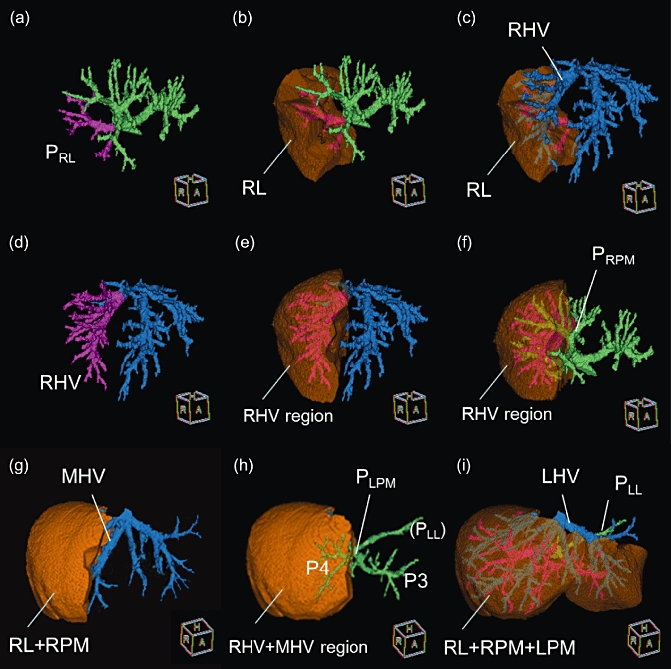
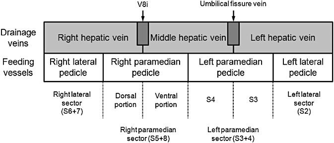
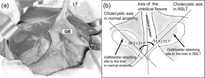
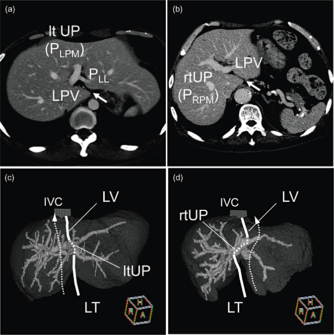
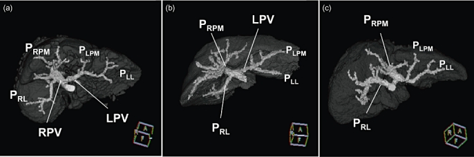
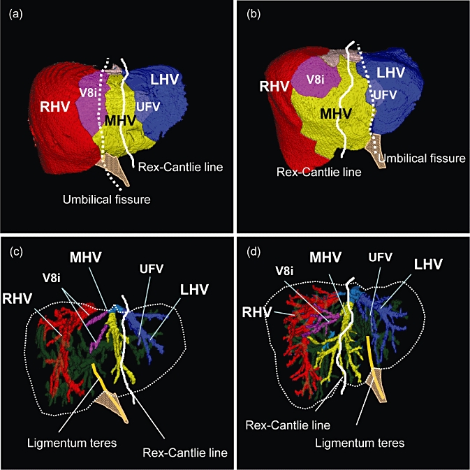
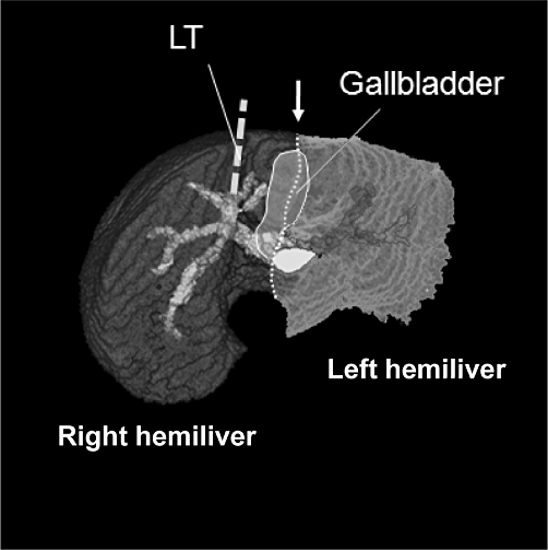
Results: Couinaud's four sectors and three hepatic veins were clearly distinguished in the liver with RSLT using 3D simulations. The ligamentum teres was connected with the right paramedian portal pedicle, and the long axis of the cystic fossa was always observed on the left of the ligamentum teres in all 35 livers. However, when the main portal scissura was visualized using 3D simulation, the gallbladder was always located on the border of either side of the hemilivers, and the malposition of the gallbladder was not confirmed.

Conclusions: Although the right-sided components of the livers are well developed as a result of the right-dominant distribution of the feeding vessels in livers with RSLT, the basic segmental structure defined by the four sectors and the three hepatic veins are as well preserved as those in the typical liver anatomy.


Figures

Figure 1
Figure 1
Step-by-step definitions of the vasculature in livers with right-sided ligamentum teres (RSLT). (a) Three-dimensional configurations of portal branches (green) and the right lateral portal pedicle (PRL) (pink). (b) Visualization of the right lateral sector. (c) When the hepatic veins (blue) are superimposed on the 3D images of right lateral sector, the right hepatic vein (RHV) is visible on the sectoral border. (d) The RHV (pink) is identified. (e) The drainage area of the RHV is then visualized. (f) The right paramedian portal pedicle (PRPM) is visible on the intersectoral plane emerging after visualizing of the RHV. (g) The MHV was confirmed after visualization of the right hemiliver (RL + RPM). RL, right lateral sector; RPM, right paramedian sector. (h) The left paramedian portal pedicle (PLPM) is visible in the umbilical fissure. (i) The left hepatic vein (LHV) was exposed on the sectoral border and the left lateral portal pedicle (PLL) can also be seen behind the LHV
Figure 2
Figure 2
Classification of liver segments according to feeding and drainage patterns. Four sectors were first defined as the regions fed by 2nd-order portal pedicles. Then, either side of the paramedian sector was further sub-divided according to the drainage veins. The right paramedian sector is divided into ‘longitudinal’ two parts: ventral and dorsal portions based on the venous drainage patterns
Figure 3
Figure 3
Relation between the umbilical portion and the gallbladder in livers with right-sided ligamentum teres (RSLT) and those with the typical liver anatomy. (a) Surgical picture of RSLT. The gallbladder (GB) is located to the left of the ligamentum teres (LT). (b) Angles between the umbilical axis and the cholecystic axis. The gallbladder was always located to the left in relation to the ligamentum teres
Figure 4
Figure 4
Characteristics of the positions of the umbilical portion and the ligamentum venosum. (a) Typical liver anatomy. The groove containing the ligamentum venosum (arrow) usually terminates where the left portal vein curves to form the umbilical portion (ltUP) ramifying with the left lateral pedicle (PLL). (b) Right-sided ligamentum teres (RSLT). The umbilical portion (rtUP) was always directed to the left towards the ligamentum venosum. The groove for the ligamentum venosum (arrow heads) was deeply cleaved reaching the bifurcation between the rtUP and the left portal vein (LPV). (c, d) The course of umbilical circulation. Intrahepatic portal veins are visualized in a 3D manner. Although the umbilical circulation course was relatively straight in the typical liver anatomy (c), that in the livers with RSLT always exhibited a ‘twisted’ course (d) when observed in 3D images
Figure 5
Figure 5
Three portal ramification patterns in livers with right-sided ligamentum teres (RSLT). (a) Bifurcation type (n = 10), (b) trifurcation type (n = 8) and (c) independent right lateral type (n = 17). PRL, right lateral portal pedicle; PRPM, right paramedian portal pedicle; PLPM, left paramedian portal pedicle; PLL, left lateral portal pedicle; LPV, left portal vein; and RPV, right portal vein
Figure 6
Figure 6
The distribution of venous drainage areas. (a, c) RSLT case, (b, d) typical liver anatomy. Each venous drainage area is represented in a different colour on the liver surface. The white line represents the simulated Rex-Cantlie line and the dotted line indicates the umbilical fissure. The umbilical fissure is located between the areas of the middle hepatic vein (MHV) and the right hepatic vein (RHV) in the livers with right-sided ligamentum teres (RSLT), while it is usually observed between the MHV and the LHV in the typical liver anatomy
Figure 7
Figure 7
Ramification patterns of the hepatic arteries in 32 right-sided ligamentum teres (RSLT) patients. The frequency of each ramification pattern is indicated on the right lower side of the respective schemes, stratified according to the portal ramification pattern. B, bifurcation type; T, trifurcation type; P, independent right lateral type. ARL, right lateral artery; ARPM, right paramedian artery; ARPM-v, ventral branch; ARPM-d, dorsal branch; ALPM, left paramedian artery; ALL, left lateral artery; PHA, proper hepatic artery; CHA, common hepatic artery; GDA, gastroduodenal artery; LGA, left gastric artery
Figure 8
Figure 8
Position of the gallbladder in relation to vascular distributions. When the intersectoral plane between each side of the hemiliver (arrow) is visualized in 3D simulation, the gallbladder was always observed on the sectoral border in 35 right-sided ligamentum teres (RSLT) cases. LT, ligamentum teres

References

    1. Nagai M, Kubota K, Kawasaki S, Takayama T, Bandai Y, Makuuchi M. Are left sided gallbladders really located on the left side? Ann Surg. 1997;225:274–280. - PMC - PubMed
    1. Maetani Y, Itoh K, Kojima N, Tabuchi T, Shibata T, Asonuma K, et al. Portal vein anomaly associated with deviation of the ligamentum teres to the right and malposition of the gallbladder. Radiology. 1998;207:723–728. - PubMed
    1. Uesaka K, Yasui K, Morimoto T, Torii A, Kodera Y, Hirai T, et al. Left-sided gallbladder with intrahepatic portal venous anomalies. J Hepatobiliary Pancreat Surg. 1995;2:425–430.
    1. Lucidarme O, Taboury J, Savier E, Cadi M, Hannoun L, Grenier PA. Fusion of the midplane with the left intersectional plane: a liver anatomical variation revisited with multidetector-row CT. Eur Radiol. 2006;16:1699–1708. - PubMed
    1. Rocca JP, Rodriguez-Davalos MI, Burke-Davis M, Marvin MR, Sheiner PA, Facciuto ME. Living-donor hepatectomy in right-sided round-ligament liver: importance of mapping the anatomy to the left medial segment. J Hepatobiliary Pancreat Surg. 2006;13:454–457. - PubMed

Publication types

MeSH terms