. 2012 Sep;16(9):2094-103.
doi: 10.1111/j.1582-4934.2011.01509.x.

Immunogenicity of allogeneic mesenchymal stem cells

Affiliations

Immunogenicity of allogeneic mesenchymal stem cells

Sabine Schu et al. J Cell Mol Med. 2012 Sep.

Abstract

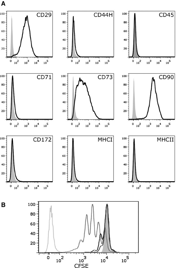
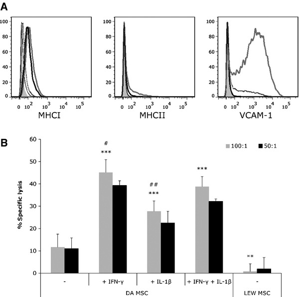
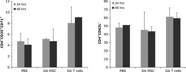
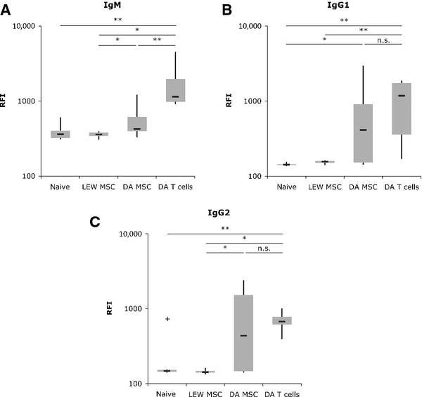
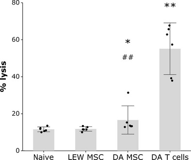
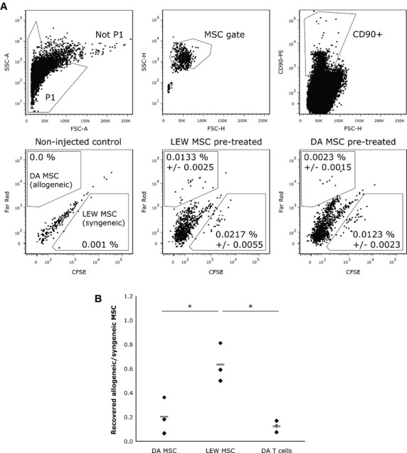
Mesenchymal stem cells (MSCs) inhibit proliferation of allogeneic T cells and express low levels of major histocompatibility complex class I (MHCI), MHCII and vascular adhesion molecule-1 (VCAM-1). We investigated whether their immunosuppressive properties and low immunophenotype protect allogeneic rat MSCs against cytotoxic lysis in vitro and result in a reduced immune response in vivo. Rat MSCs were partially protected against alloantigen-specific cytotoxic T cells in vitro. However, after treatment with IFN-γ and IL-1β, MSCs upregulated MHCI, MHCII and VCAM-1, and cytotoxic lysis was significantly increased. In vivo, allogeneic T cells but not allogeneic MSCs induced upregulation of the activation markers CD25 and CD71 as well as downregulation of CD62L on CD4(+) T cells from recipient rats. However, intravenous injection of allo-MSCs in rats led to the formation of alloantibodies with the capacity to facilitate complement-mediated lysis, although IgM levels were markedly decreased compared with animals that received T cells. The allo-MSC induced immune response was sufficient to lead to significantly reduced survival of subsequently injected allo-MSCs. Interestingly, no increased immunogenicity of IFN-γ stimulated allo-MSCs was observed in vivo. Both the loss of protection against cytotoxic lysis under inflammatory conditions and the induction of complement-activating antibodies will likely impact the utility of allogeneic MSCs for therapeutic applications.


Figures

Fig 1
Fig 1
Characterization of rat mesenchymal stem cells (MSCs). (A) rMSCs are CD29+, CD73+, CD90+, and major histocompatibility complex class I (MHCI), MHCII, CD44H, CD45, CD71, CD172 low or negative. Shown are FACS histograms of Dark Agouti (DA) rMSCs (passage 5) stained with antibodies against surface markers as indicated (black) or with appropriate isotype controls (grey). (B) CFSE-labelled T cells were polyclonally stimulated with anti-CD3/anti-CD28-labelled beads in the absence (medium grey line) or presence of MSCs in different ratios (black line: 1:10; dark grey line: 1:5). CFSE fluorescence was analysed on day 3. Unstimulated T cells (filled grey line) and unstained T cells (light grey line) served as controls. Shown is a representative experiment of >3.
Fig 2
Fig 2
Pretreatment with inflammatory cytokines leads to upregulation of major histocompatibility complex class I (MHCI), MHCII and vascular adhesion molecule-1 (VCAM-1), and renders allogeneic rat mesenchymal stem cells (rMSCs) susceptible to cytotoxic lysis by alloantigen-specific T cells. (A) MSCs were treated with 100 U/ml IFN-γ, IL-1β or IFN-γ + IL-1β for 24 hr and analysed for MHCI, MHCII and VCAM-1 expression by FACS. IFN-γ (thick black line) and IFN-γ + IL-1β (grey line) induced upregulation of MHCI and MHCII, while MSCs stimulated with IL-1β alone (dotted line) did not change MHCII expression and only slightly increased MHCI expression compared to untreated MSCs (thin black line). VCAM-1 expression was induced by IL-1β and IFN-γ + IL-1β, but not by IFN-γ alone. Isotype controls are shown in filled grey (representative experiment from >3). (B) Alloantigen-specific cytotoxic T cells (CTLs) were generated in a mixed lymphocyte culture of LEW and γ-irradiated DA T cells. Syngeneic LEW or allogeneic DA rMSCs, either untreated or pretreated with 100 U/ml IFN-γ, IL-1β or IFN-γ + IL-1β for 24 hr, were stained with the fluorescent dye calcein and cocultured with alloantigen-specific CTLs in an effector to target ratio of 100:1 or 50:1 for 4 hr. MSCs that are lysed by CTLs release calcein into the cell culture supernatant and fluorescence of the supernatant is proportional to the amount of cells lysed. Percentage of specific lysis is calculated in relation to spontaneous release of calcein of MSCs in medium alone and maximum release of calcein by Triton-X treated MSCs. Shown is a representative experiment with means of five replicates ± S.D. (**P < 0.01; ***P < 0.001 compared to untreated DA MSC; #P < 0.05; ##P < 0.01 compared to DA MSC pretreated with IFN-γ + IL-1β; student's t-test).
Fig 3
Fig 3
Allogeneic mesenchymal stem cells (MSCs) do not induce markers of T cell activation. LEW rats were injected with 5 × 10⁶ DA MSCs, DA T cells or PBS. Spleens were harvested 24 or 48 hr after injection and expression of early activation markers was analysed. Cells were stained with anti-CD4, anti-CD25, and anti-CD71 (left) or anti-CD4 and anti-CD62L (right), or appropriate isotype controls. Shown are mean percentages of positive cells ± S.D. (n = 2–3).
Fig 4
Fig 4
Allogeneic mesenchymal stem cells (MSCs) elicit an antibody response. LEW rats were injected with 1 × 10⁶ syngeneic or allogeneic (DA) MSCs or allogeneic T cells. Serum was harvested 2–3 weeks after injection. Splenocytes of allogeneic DA rats were incubated with serum aliquots and, consequently, bound alloantigen-specific antibodies were stained with anti-rat IgM (A), anti-rat IgG1 (B) or anti-rat IgG2 (C) antibodies. B cells were excluded from analysis of IgM by counterstaining with CD45RA (n = 6 animals/group; *P < 0.05; **P < 0.01; n.s.: not significant; non-parametric Mann–Whitney test).
Fig 5
Fig 5
Allogeneic mesenchymal stem cell (MSC)-induced antibodies mediate significant albeit reduced complement activation. LEW rats were injected with 1 × 10⁶ syngeneic or allogeneic MSCs or allogeneic T cells. Serum was harvested 2–3 weeks after injection. Splenocytes of allogeneic DA rats were incubated with serum (diluted 1:10) and treated with baby rabbit complement (diluted 1:10). Splenocytes were stained with PI to detect complement-mediated lysis. The grey bars show the means and 95% confidence intervals of serums from six animals per group tested and the black dots show means of triplicates from serum of individual animals. *P < 0.05, **P < 0.01 compared to LEW MSC and naïve; ##P < 0.01 compared to DATc (non-parametric Mann–Whitney test).
Fig 6
Fig 6
Reduced survival of allogeneic mesenchymal stem cells (MSCs) in vivo. LEW rats were injected with 3.5 × 10⁶ syngeneic or allogeneic MSCs or allogeneic T cells (three animals per group). After 2 weeks, all groups received 2.5 × 10⁶ CFSE-stained LEW MSCs and Far Red-stained DA MSCs (ratio 1:1). Lungs were harvested 24 hr after injection. Mononuclear cells were enriched using a Ficoll gradient and stained with anti-rat CD90-PE. (A) Gating strategy: Not P1 → MSC gate → CD90+ (top row). Bottom row: Shown are representative dot plots of CD90+ cells recovered from lungs of non-injected controls, LEW MSC pre-treated and DA MSC pre-treated rats (left to right). The percentages given are percentage of events ± S.D. in the MSC gate that are CD90+ CFSE+ (LEW MSCs) or CD90+ Far Red+ double positive (DA MSCs), respectively. (B) Ratio of allogeneic MSCs (CD90+ Far Red+) to syngeneic MSCs (CD90+ CFSE+) in LEW rats pre-treated with DA MSCs or LEW MSCs. Rats pre-treated with DA T cells were used as positive controls. Each black diamond represents an individual animal (n = 3/group), the grey lines show the mean (*P < 0.05; non-parametric Mann–Whitney test).

References

    1. Sensebe L, Bourin P. Mesenchymal stem cells for therapeutic purposes. Transplantation. 2009;87:S49–53. - PubMed
    1. Aggarwal S, Pittenger MF. Human mesenchymal stem cells modulate allogeneic immune cell responses. Blood. 2005;105:1815–22. - PubMed
    1. Bernardo ME, Locatelli F, Fibbe WE. Mesenchymal stromal cells. Ann N Y Acad Sci. 2009;1176:101–17. - PubMed
    1. Morigi M, Imberti B, Zoja C, et al. Mesenchymal stem cells are renotropic, helping to repair the kidney and improve function in acute renal failure. J Am Soc Nephrol. 2004;15:1794–804. - PubMed
    1. Togel F, Hu Z, Weiss K, et al. Administered mesenchymal stem cells protect against ischemic acute renal failure through differentiation-independent mechanisms. Am J Physiol Renal Physiol. 2005;289:F31–42. - PubMed

Publication types

MeSH terms