. 2012 Jan-Feb;20(1):38-49.
doi: 10.1111/j.1524-475X.2011.00748.x. Epub 2011 Dec 8.

Tumor necrosis factor-alpha (TNF-α) is a therapeutic target for impaired cutaneous wound healing

Affiliations

Tumor necrosis factor-alpha (TNF-α) is a therapeutic target for impaired cutaneous wound healing

Gillian S Ashcroft et al. Wound Repair Regen. 2012 Jan-Feb.

Abstract

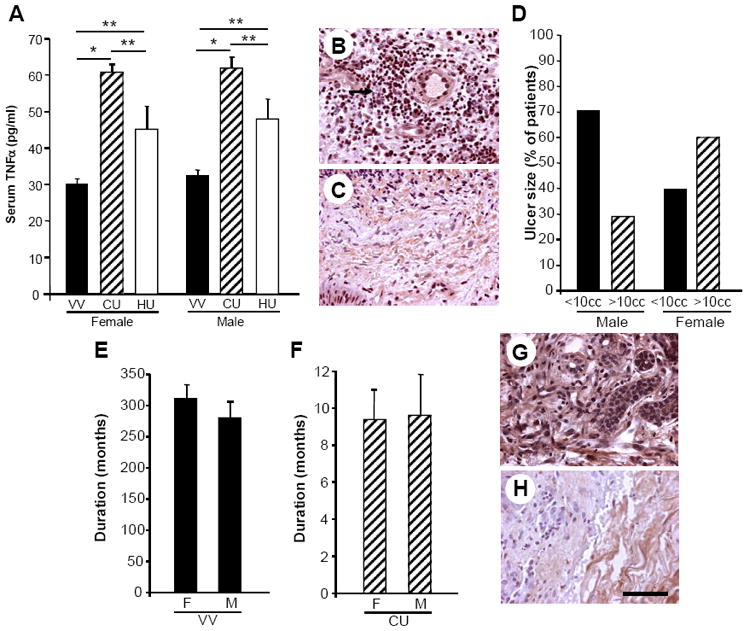
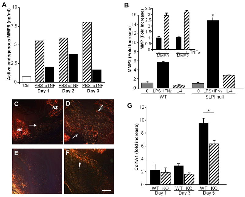
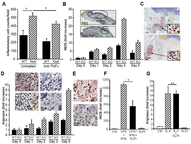
Impaired wound healing states lead to substantial morbidity and cost with treatment resulting in an expenditure of billions of dollars per annum in the U.S. alone. Both chronic wounds and impaired acute wounds are characterized by excessive inflammation, enhanced proteolysis, and reduced matrix deposition. These confounding factors are exacerbated in the elderly, in part, as we report here, related to increased local and systemic tumor necrosis factor-alpha (TNF-α) levels. Moreover, we have used a secretory leukocyte protease inhibitor (SLPI) null mouse model of severely impaired wound healing and excessive inflammation, comparable to age-related delayed human healing, to demonstrate that topical application of anti-TNF-α neutralizing antibodies blunts leukocyte recruitment and NFκB activation, alters the balance between M1 and M2 macrophages, and accelerates wound healing. Following antagonism of TNF-α, matrix synthesis is enhanced, associated with suppression of both inflammatory parameters and NFκB binding activity. Our data suggest that inhibiting TNF-α is a critical event in reversing the severely impaired healing response associated with the absence of SLPI, and may be applicable to prophylaxis and/or treatment of impaired wound healing states in humans.


Conflict of interest statement

The authors have no conflicting financial interests.

Figures

Figure 1
Figure 1. TNFα levels are increased locally and systemically in human subjects predisposed to impaired healing
A) Systemic TNFα levels, determined by ELISA on serum samples, are significantly increased in subjects who have previously suffered from, or who currently have, chronic venous ulceration (CU) compared to age-matched controls with underlying pathology (i.e. venous hypertension/varicose veins, VV), but not unresolved inflammation. Female VV n=43; Female CU n=25; Male VV n=38; Male CU n=17. Data represent mean +/- SEM *p < 0.0001. Serum samples were also collected from individuals whose venous ulcer had healed (healed ulcer, HU) (n = 6 male, 9 female) and serum levels of TNFα were significantly (**p < 0.05) higher in subjects with an active venous ulcer (CU) compared with those with healed ulcers (HU). Subjects with healed ulcers (HU) also showed significantly (**p < 0.05) increased systemic TNFα levels compared with those measured in the control population (VV). B) Immunohistochemical analysis revealed local TNFα levels to be high in initial chronic ulcer biopsies (arrow indicates inflammatory cells staining positively for TNFα) and decrease as chronic venous ulcers heal (C). Representative patient biopsy images before and after healing, n=4. D) Ulcer size in male (n=17) and female (n=25) subjects represented as percentage of subjects with ulcers greater or less than 10cc. E) Duration of varicose veins in male and female subjects in months. F) Duration of venous ulcers in male and female subjects in months. G) Acute day 7 wound biopsies from the upper inner arm (site distant from chronic ulcer) show increased TNFα immunostaining in chronic ulcer patients compared to healthy controls (H). Representative images, n=4. Bar = 40 μm.
Figure 2
Figure 2. Increased TNFα in murine model of delayed healing
A) Thioglycollate-induced peritoneal exudate macrophages (pooled from 3-5 mice) from SLPI null and WT mice were treated with a TLR4 agonist LPS (100ng/ml) for 1 hr and the cells processed for RT-PCR analysis. TLR4 stimulation of SLPI null macrophages induced higher levels of TNFα than for WT macrophages. N=2. B) By conventional RT-PCR, TNFα expression is minimal in control skin and enhanced in wounds from SLPI null mice compared to WT wounds at day 3 post-wounding treated with PBS vehicle. Local treatment with recombinant SLPI reduced TNFα expression in the SLPI null mice wounds to near levels seen in WT and resulted in accelerated healing. N=6 animals. *p < 0.05 comparing null control with null PBS-treated and ** p <0.05 for null PBS-treated vs. null SLPI-treated (black bars). Inset: Representative experiment showing conventional RT-PCR for TNFα expression in control skin and wounds treated locally with PBS vehicle or with SLPI. C,D) Increased intensity of immunostaining for TNFα in day 3 SLPI null skin wounds (D) relative to WT mice (C).
Figure 3
Figure 3. TNFα as a target of SLPI and accelerated healing by TNFα neutralization
A) Pooled WT peritoneal macrophages were stimulated with LPS in culture in the presence or absence of SLPI (10 μg/ml added 30min prior to LPS) which significantly suppressed TNFα expression determined by real time PCR 1hr after stimulation with LPS. *p<0.05. Representative experiment, N=2. B, C) Exogenous TNFα added to peritoneal macrophages induces significant TNFα expression (B) in parallel with augmented ADAM17/TACE expression (C) which activates TNFα to enhance a cycle of pro-inflammatory activity. *p<0.05. D, E) Local administration of anti-TNFα reverses delayed healing phenotype in SLPI null mice. At the macroscopic level, anti-TNFα antibody treatment (αTNFα) accelerates healing in SLPI null mouse (D) to a greater extent than in the WT (E) (day 3 post-wounding shown). F) Anti-TNFα antibodies applied at the time of wounding significantly reduce cross-sectional wound areas in SLPI null mice compared to control (PBS treated) wounds at day 3 post-wounding. Areas= 105 mm2. Data represent mean +/- SEM * P < 0.05. N=6. G,H) Masson’s Trichrome staining illustrates a marked reduction of wound size in representative SLPI null mouse treated with anti-TNFα antibodies (G) compared to PBS (H). Arrows demarcate wound margins. Bar (H) = 300 μm (G-H).
Figure 4
Figure 4. MMP is reduced and deposition of matrix protein is increased by anti-TNFα treatment
A) Supernatants from pooled and cultured SLPI null wound tissues at days 1-3 post wounding were tested for MMP9 activity by ELISA. Increased levels of active MMP9 were seen in the PBS-treated wounds cultured ex vivo compared to control skin. Treatment of wounds in vivo with anti-TNFα antibodies resulted in reduced levels of active MMP9 generated in parallel cultures from wounds on day 1, 2 or 3. B) Inducible macrophage MMP expression (MMP2 shown) is consistent with classically activated (M1), rather than alternatively activated macrophages (IL-4, M2) and is higher in SLPI null relative to WT peritoneal macrophages. null vs WT *p<0.01. Inset: TNFα independently stimulates MMP2 and MMP9 expression in WT (shown) and null macrophages as determined by RT-PCR. C-F) Picrosirius red staining of tissues at day 3 post-wounding illustrates increased collagen deposition (arrows) in anti-TNFα treated wounds compared to untreated control: C) WT day 3 post-wounding. D) WT treated with anti-TNFα day 3. E) SLPI null day 3 wound. F) SLPI null wound treated with anti-TNFα. NS= normal skin adjacent to wound. Bar (F) = 150 μm (C-F). G) Wound tissues at indicated intervals post wounding were evaluated by RT-PCR for type I collagen (Col1A1) gene expression. *p<0.05
Figure 5
Figure 5. Increased inflammatory cell infiltrate reflecting M1 bias in SLPI deficient mice
A) Inflammatory cell quantification demonstrates significantly increased inflammatory cells per unit area in SLPI null compared to wild-type (WT) wounds at day 3, as described (1). Treatment with anti-TNFα significantly reduced inflammatory infiltrate. Data represent mean+/- SEM *P<0.05. B) Wound tissues were assessed by RT-PCR for expression levels of iNOS as a marker for M1 macrophages, which were elevated and sustained in null relative to WT wounds. Inset: PBS treated(top) and anti-TNFα treated wounds showing reduced wound size with treatment. Day 3, original magnification 5X. C) Reduced iNOS staining in anti-TNFα treated wounds(bottom) relative to untreated(top) wounds, consistent with a decrease in M1 pro-inflammatory cells. Insets: Mac2 staining on serial sections showing macrophages within infiltrated region of iNOS staining. Representative staining, n=4. D) Arginase as detected by RT-PCR and protein(inset) reveal increasing expression of arginase in SLPI null wounds over time as compared to parallel wounds in WT mice. Inset: With TNFα inhibition, arginase expressing inflammatory cells by immunohistochemistry decrease as inflammation resolves and fibroblasts appear more numerous in SLPI null mice(top two panels of inset: left panel day 3 wound; right panel day 3 post anti-TNFα) and in WT (bottom two panels of inset: left panel day 3 wound; right panel day 3 post anti-TNFα). E) Day 3 wound serial tissue sections stained for arginase (top panel) and Mac2 (bottom) identify areas enriched in macrophages that are positive for detection of arginase, original magnification 40X. F) Thioglycollate-induced peritoneal macrophages were treated in culture with IFNγ (10ng/ml) and LPS(100ng/ml) to induce M1 phenotype and expression of iNOS monitored in presence or absence of SLPI(10μg/ml). SLPI inhibited M1 iNOS expression, *p=0.03. G) Thioglycollate-induced macrophages were treated with IL-4(10ng/ml) to induce M2 phenotype and arginase expression determined by RT-PCR in presence or absence of SLPI(10μg/ml). SLPI did not suppress arginase (n=3). **P=NS.
Figure 6
Figure 6. Enhanced NFκB binding activity in the absence of SLPI is reduced in anti-TNFα treated wounds
A) Peritoneal macrophages from WT and SLPI null mice were stimulated with LPS for indicated intervals and NFκB activation monitored by Western blot (p65) showing increased activation of this transcription factor in the absence of SLPI. In parallel, by Western blot, IκB, high in unstimulated macrophages, is degraded within minutes after exposure to LPS, and then the levels return to normal in the WT, whereas a slower recovery of the inhibitor occurs in the absence of SLPI. B) NFκB DNA binding is increased in nuclear extracts from SLPI null (KO) normal skin compared to wild-type (WT) normal skin tissues. Increased activity at day 1 post-wounding, particularly in the SLPI null wounds was reduced by anti-TNFα treatment (+). (− = PBS, no anti-TNFα). N = 6. C, D) NFκB phospho-p65 staining in KO (C) and WT (D) wounds at day 3 showing enhanced numbers of inflammatory cells and increased p65 staining in SLPI null wounds. Inset: Higher magnification (original magnification 40X) showing nuclear staining.

References

    1. Ashcroft GS, Greenwell-Wild T, Horan MA, Wahl SM, Ferguson MW. Topical estrogen accelerates cutaneous wound healing in aged humans associated with an altered inflammatory response. Am J Pathol. 1999;155(4):1137–46. - PMC - PubMed
    1. Schafer M, Werner S. Cancer as an overhealing wound: an old hypothesis revisited. Nat Rev Mol Cell Biol. 2008;9(8):628–38. - PubMed
    1. Song X, Zeng L, Jin W, Thompson J, Mizel DE, Lei K, Billinghurst RC, Poole AR, Wahl SM. Secretory leukocyte protease inhibitor suppresses the inflammation and joint damage of bacterial cell wall-induced arthritis. J Exp Med. 1999;190(4):535–42. - PMC - PubMed
    1. Feldmann M, Williams RO, Paleolog E. What have we learnt from targeted anti-TNF therapy? Ann Rheum Dis. 2010;69(Suppl 1):i97–9. - PubMed
    1. Chan JM, Villarreal G, Jin WW, Stepan T, Burstein H, Wahl SM. Intraarticular gene transfer of TNFR:Fc suppresses experimental arthritis with reduced systemic distribution of the gene product. Mol Ther. 2002;6(6):727–36. - PubMed

Publication types

MeSH terms