. 2011 Dec 12:5:575.
doi: 10.1186/1752-1947-5-575.

Diffuse bronchiolitis pattern on a computed tomography scan as a presentation of pulmonary tumor thrombotic microangiopathy: a case report

Affiliations

Diffuse bronchiolitis pattern on a computed tomography scan as a presentation of pulmonary tumor thrombotic microangiopathy: a case report

Marcos Duarte Guimarães et al. J Med Case Rep. .

Abstract

Introduction: Pulmonary tumor thrombotic microangiopathy is a rare complication of malignant diseases. The diagnosis is extremely difficult and is most often performed after death. Invariably, patients develop acute pulmonary hypertension causing right heart failure, shortness of breath and death in a few days. We describe the clinical and radiological findings of a patient who presented with this complication.

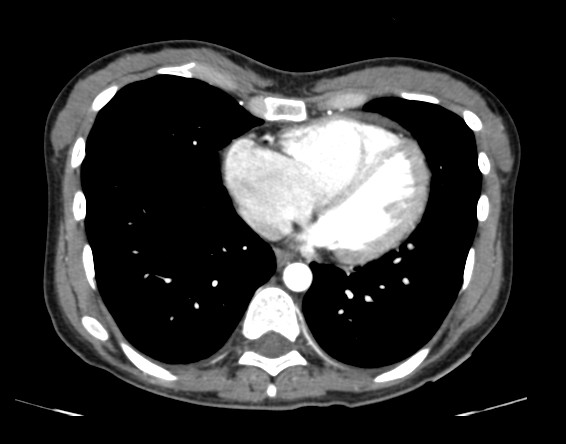
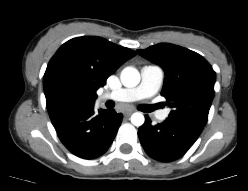
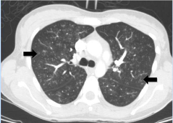
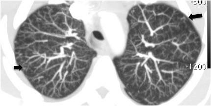
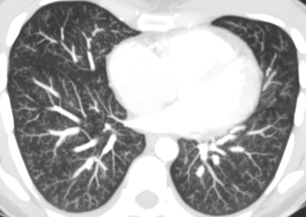
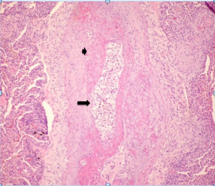
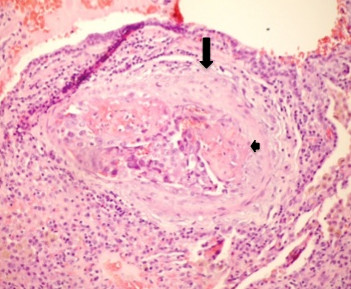
Case presentation: A 28-year-old Caucasian woman with a previous history of pelvic tumor resection two months previously, suggestive of metastatic adenocarcinoma, presented with intense shortness of breath. A computed tomography scan showed signs of acute cor pulmonale and diffuse nodular opacities associated with a tree-in-bud pattern disseminated through her lungs, suggestive of bronchiolitis. Our patient's condition worsened and she underwent a surgical biopsy. Pathologic analysis of the biopsied specimens revealed pulmonary tumor thrombotic microangiopathy. Our patient's tumor evolved from a gastric origin (Krukenberg tumor). She underwent progressive clinical deterioration and died less than 24 hours after the biopsy. None of the cases described previously in the literature had diffuse centrilobular nodular opacities associated with a tree-in-bud pattern disseminated through the lungs, as in our case.

Conclusion: Pulmonary tumor thrombotic microangiopathy should be considered in cancer patients with rapidly progressing dyspnea, chest computed tomography findings compatible with pulmonary hypertension and typical findings of inflammatory bronchiolitis.


Figures

Figure 1
Figure 1
CT axial series with medastinal window after infusion of intravenous contrast, showing increase in the size of the right atrium and right ventricle. The right ventricular to left ventricular ratio is approximately 1.8.
Figure 2
Figure 2
CT axial series with medastinal window after infusion of intravenous contrast showing slight increase in the caliber of the pulmonary artery measuring about 30 mm.
Figure 3
Figure 3
High resolution CT scan shows diffuse pulmonary involvement with ill-defined centrilobular nodules and branching lines (arrows), which correspond to tree-in-bud appearance.
Figure 4
Figure 4
CT axial series of the upper lobes with maximum intensity projection showing diffuse centrilobular nodular opacities with ground-glass attenuation associated with tree-in-bud pattern.
Figure 5
Figure 5
CT axial series of the lower lobes with maximum intensity projection showing diffuse centrilobular nodular opacities with ground-glass attenuation associated with tree-in-bud pattern.
Figure 6
Figure 6
Photomicrograph of histopathologic specimen shows complete arteriolar occlusion by tumor cells (arrow) and fibrointimal proliferation (small arrow), surrounded by lung tissue with collapsed alveoli. (Original magnification ×400; hematoxylin-eosin stain.)
Figure 7
Figure 7
Photomicrograph of histopathologic specimen shows fibrointimal proliferation (arrow) and presence of thrombi (small arrow) surrounded by tumor cells in centrilobular arteriole. (Original magnification ×400; hematoxylin-eosin stain.)

Similar articles

Cited by

References

    1. Shih HM, Lin CC, Shiao YW. Pulmonary tumor thrombotic microangiopathy. Am J Emerg Med. 2011;29(2):241.e3–241.e4. doi: 10.1016/j.ajem.2010.03.004. - DOI - PubMed
    1. Yokomine T, Hirakawa H, Ozawa E, Shibata K, Nakayama T. Pulmonary thrombotic microangiopathy caused by gastric carcinoma. J Clin Pathol. 2010;63(4):367–369. doi: 10.1136/jcp.2010.075739. - DOI - PMC - PubMed
    1. von Herbay A, Waldherr R, Otto HF. Pulmonary tumor thrombotic microangiopathy with pulmonary hypertension. Cancer. 1990;66:587–592. doi: 10.1002/1097-0142(19900801)66:3<587::AID-CNCR2820660330>3.0.CO;2-J. - DOI - PubMed
    1. Sakashita N, Yokose C, Fujii K, Matsumoto M, Ohnishi K, Takeya M. Pulmonary tumor thrombotic microangiopathy resulting from metastatic signet ring cell carcinoma of the stomach. Pathol Int. 2007;57(6):383–387. doi: 10.1111/j.1440-1827.2007.02111.x. - DOI - PubMed
    1. Malani AK, Gupta C, Kutty AV, Betlej T. Pulmonary tumor thrombotic microangiopathy from metastatic gallbladder carcinoma: an unusual cause of severe pulmonary hypertension. Dig Dis Sci. 2007;52(2):555–557. doi: 10.1007/s10620-006-9232-z. - DOI - PubMed

LinkOut - more resources