

Vitreous inflammatory factors and serous retinal detachment in central retinal vein occlusion: a case control series
- PMID: 22152024
- PMCID: PMC3253063
- DOI: 10.1186/1476-9255-8-38
Vitreous inflammatory factors and serous retinal detachment in central retinal vein occlusion: a case control series
Abstract
Background: This study investigated whether the vitreous fluid levels of soluble vascular endothelial growth factor receptor-2 (sVEGFR-2), pigment epithelium-derived factor (PEDF), and soluble intercellular adhesion molecule 1 (sICAM-1) were associated with the occurrence of serous retinal detachment (SRD) in patients with central retinal vein occlusion (CRVO).
Methods: We recruited 33 patients with CRVO and macular edema, as well as 18 controls with nonischemic ocular diseases. Eighteen of the 33 patients with CRVO showed SRD on optical coherence tomography of the macula (defined as subretinal accumulation of fluid with low reflectivity), while the other 15 patients only had cystoid macular edema (CME, defined as hyporeflective intraretinal cavities). Retinal ischemia was evaluated by measuring the area of capillary non-perfusion using fluorescein angiography and the public domain Scion Image program, while central macular thickness (CMT) was examined by optical coherence tomography. Vitreous fluid samples were obtained during pars plana vitrectomy and levels of the target molecules were measured by enzyme-linked immunosorbent assay.
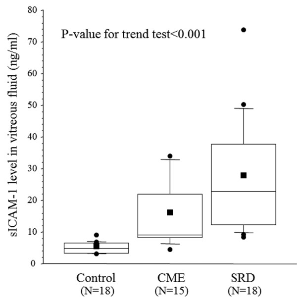
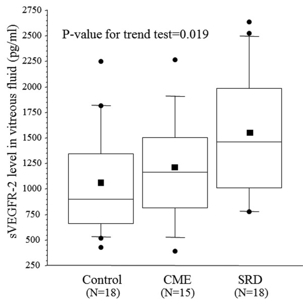
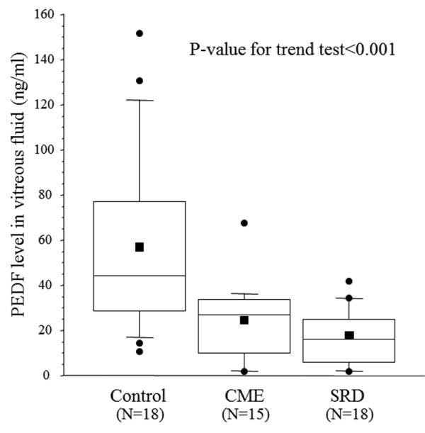
Results: Ischemia was significantly more common in the SRD group (17/18 patients) than in the CME group (5/15 patients) (P < 0.001). The vitreous fluid level of sICAM-1 increased significantly across the three groups from the control group (4.98 ± 1.73 ng/ml) to the CME group (15.4 ± 10.1 ng/ml) and the SRD group (27.1 ± 17.7 ng/ml) (ptrend< 0.001). The vitreous fluid level of sVEGFR-2 also showed a significant increase across the three groups (1083 ± 541 pg/ml, 1181 ± 522 pg/ml, and 1535 ± 617 pg/ml, respectively, ptrend = 0.019). On the other hand, the vitreous fluid level of PEDF showed a significant decrease across the three groups (56.4 ± 40.0 ng/ml, 24.3 ± 17.3 ng/ml, and 16.4 ± 12.6 ng/ml, respectively, ptrend< 0.001).
Conclusions: Higher levels of inflammatory factors (sICAM-1 and sVEGFR-2) and lower levels of anti-inflammatory PEDF were observed in macular edema patients with SRD, suggesting that inflammation plays a key role in determining the severity of CRVO.
Figures
Similar articles
-
Vitreous inflammatory factors and serous macular detachment in branch retinal vein occlusion.Retina. 2012 Jan;32(1):86-91. doi: 10.1097/IAE.0b013e31821801de. Retina. 2012. PMID: 21866074
-
Vitreous inflammatory factors in macular edema with central retinal vein occlusion.Jpn J Ophthalmol. 2011 May;55(3):248-255. doi: 10.1007/s10384-011-0016-4. Epub 2011 May 3. Jpn J Ophthalmol. 2011. PMID: 21538003
-
Aqueous vascular endothelial growth factor levels are associated with serous macular detachment secondary to branch retinal vein occlusion.Retina. 2010 Feb;30(2):281-6. doi: 10.1097/IAE.0b013e3181b9f153. Retina. 2010. PMID: 19881397
-
Soluble vascular endothelial growth factor receptor-2 and inflammatory factors in macular edema with branch retinal vein occlusion.Am J Ophthalmol. 2011 Oct;152(4):669-677.e1. doi: 10.1016/j.ajo.2011.04.006. Epub 2011 Jul 2. Am J Ophthalmol. 2011. PMID: 21726846
-
Vitreous levels of interleukin-6 and vascular endothelial growth factor in macular edema with central retinal vein occlusion.Ophthalmology. 2009 Jan;116(1):87-93. doi: 10.1016/j.ophtha.2008.09.034. Ophthalmology. 2009. PMID: 19118700
Cited by
-
Increased expression of angiogenic and inflammatory proteins in the vitreous of patients with ischemic central retinal vein occlusion.PLoS One. 2015 May 15;10(5):e0126859. doi: 10.1371/journal.pone.0126859. eCollection 2015. PLoS One. 2015. PMID: 25978399 Free PMC article.
-
Biochemical and microstructural determinants of the development of serous retinal detachment secondary to retinal vein occlusion.Heliyon. 2023 Dec 16;10(1):e23716. doi: 10.1016/j.heliyon.2023.e23716. eCollection 2024 Jan 15. Heliyon. 2023. PMID: 38187225 Free PMC article.
-
Atypical Presentation of Vitreous Inflammation in a Patient With Hypertensive Retinopathy.J Vitreoretin Dis. 2024 Jul 27;8(5):614-617. doi: 10.1177/24741264241264361. eCollection 2024 Sep-Oct. J Vitreoretin Dis. 2024. PMID: 39345867 Free PMC article.
-
Prevalence of Serous Macular Detachment in Recurrent Macular Edema Secondary to Retinal Vein Occlusion.Turk J Ophthalmol. 2022 Aug 25;52(4):276-280. doi: 10.4274/tjo.galenos.2021.02582. Turk J Ophthalmol. 2022. PMID: 36017487 Free PMC article.
-
Pigment epithelium-derived factor engineered to increase glycosaminoglycan affinity while maintaining bioactivity.Biochem Biophys Res Commun. 2022 May 21;605:148-153. doi: 10.1016/j.bbrc.2022.03.079. Epub 2022 Mar 17. Biochem Biophys Res Commun. 2022. PMID: 35334413 Free PMC article.
References
-
- Hayreh SS. Classification of central retinal vein occlusion. Ophthalmology. 1983;90:458–474. - PubMed
LinkOut - more resources
Full Text Sources
Miscellaneous
