

Plasmacytoid myoepithelioma of minor salivary glands: report of case with emphasis in the immunohistochemical findings
- PMID: 22152025
- PMCID: PMC3285037
- DOI: 10.1186/1746-160X-7-24
Plasmacytoid myoepithelioma of minor salivary glands: report of case with emphasis in the immunohistochemical findings
Abstract
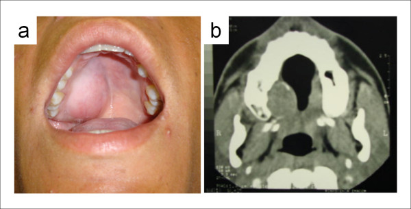
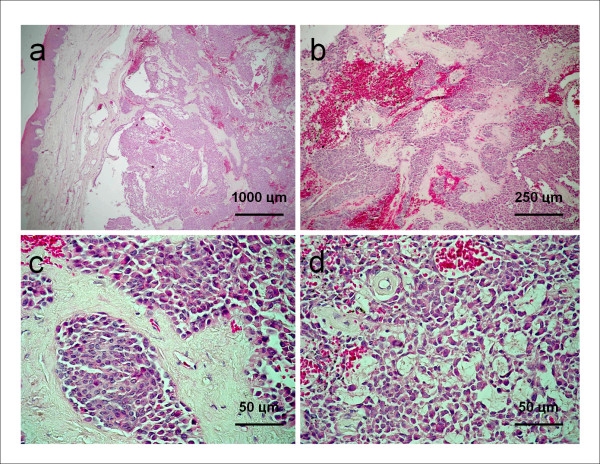
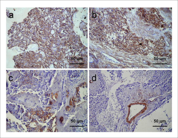
Myoepithelioma is a rare benign tumor of the salivary glands and is usually seen in the parotid gland and the minor salivary glands. It was once considered to be a type of pleomorphic adenoma (PA), but myoepitheliomas are today believed to be relatively aggressive tumors. Myoepitheliomas are most common in young adults between the ages of 30 and 50 and there are very few cases reported in individuals less than 18 years of age. We report a case of myoepithelioma located in the hard palate in a 15-year-old Brazilian male. The tumor was composed of plasmacytoid myoepithelial cells. An analysis of the immunohistochemical profile of the tumor cells showed positivity for vimentin, S-100 protein, and glial fibrillary acidic protein (GFAP), but not for smooth muscle actin (α-SMA) and cytokeratin 14 (CK14). We report this case because of the rarity of this tumor, especially in adolescents. We also discuss the histological parameters of the differential diagnosis of this tumor as well as its immunohistochemical profile.
Figures
Similar articles
-
Myoepithelial tumors of salivary glands: a clinicopathologic, immunohistochemical, ultrastructural, and flow-cytometric study.Semin Diagn Pathol. 1996 May;13(2):138-47. Semin Diagn Pathol. 1996. PMID: 8734420
-
Diagnostic criteria for neoplastic myoepithelial cells in pleomorphic adenomas and myoepitheliomas. Immunocytochemical detection of muscle-specific actin, cytokeratin 14, vimentin, and glial fibrillary acidic protein.Oral Surg Oral Med Oral Pathol Oral Radiol Endod. 1995 Mar;79(3):330-41. doi: 10.1016/s1079-2104(05)80227-6. Oral Surg Oral Med Oral Pathol Oral Radiol Endod. 1995. PMID: 7542546
-
Myoepitheliomas and myoepithelial adenomas of salivary gland origin. Immunohistochemical evaluation of filament proteins, S-100 alpha and beta, glial fibrillary acidic proteins, neuron-specific enolase, and lactoferrin.Pathol Res Pract. 1989 Feb;184(2):168-78. Pathol Res Pract. 1989. PMID: 2540482
-
Myoepithelial carcinoma of intraoral minor salivary glands: a clinicopathological study of 7 cases and review of the literature.Oral Surg Oral Med Oral Pathol Oral Radiol Endod. 2010 Jul;110(1):85-93. doi: 10.1016/j.tripleo.2010.02.023. Epub 2010 May 21. Oral Surg Oral Med Oral Pathol Oral Radiol Endod. 2010. PMID: 20488733 Review.
-
Plasmacytoid myoepithelioma of palate: three rare cases and literature review.J Laryngol Otol. 2007 Sep;121(9):e13. doi: 10.1017/S002221510700000X. Epub 2007 Jul 19. J Laryngol Otol. 2007. PMID: 17640425 Review.
Cited by
-
Myoepithelial Carcinoma Arising in a Plasmacytoid Myoepithelioma of the Parotid Gland Synchronized with Melanoma: A Case Report and Review of the Literature.Case Rep Oncol. 2021 Mar 1;14(1):173-183. doi: 10.1159/000510937. eCollection 2021 Jan-Apr. Case Rep Oncol. 2021. PMID: 33776700 Free PMC article.
-
Innervation of submandibular and sublingual glands in elderly donated cadavers: a preliminary histological study of differences in nerve morphology between mucous and serous acini.Anat Cell Biol. 2015 Mar;48(1):36-43. doi: 10.5115/acb.2015.48.1.36. Epub 2015 Mar 20. Anat Cell Biol. 2015. PMID: 25806120 Free PMC article.
-
Unusual neoplasm on the hard palate of a child: a case report.J Med Case Rep. 2017 Jun 2;11(1):149. doi: 10.1186/s13256-017-1321-4. J Med Case Rep. 2017. PMID: 28577557 Free PMC article.
-
Epithelioid osteoblastoma of maxilla: A rare and aggressive variant of a benign neoplasm at an uncommon site.J Oral Maxillofac Pathol. 2016 May-Aug;20(2):332. doi: 10.4103/0973-029X.185923. J Oral Maxillofac Pathol. 2016. PMID: 27601840 Free PMC article.
-
Heterogeneity of glandular cells in the human salivary glands: an immunohistochemical study using elderly adult and fetal specimens.Anat Cell Biol. 2013 Jun;46(2):101-12. doi: 10.5115/acb.2013.46.2.101. Epub 2013 Jun 30. Anat Cell Biol. 2013. PMID: 23869257 Free PMC article.
References
-
- Sheldon WH. So-called mixed tumors of the salivary glands. Arch Pathol. 1943;35:1–20.
-
- Cuadra ZF, Quezada RD, Tapia VJL, Paez VC, Gaitán CLA. Plasmacytoid myoepithelioma of the palate. Report of one case and review of the literature. Med Oral Patol Oral Cir Bucal. 2007;12:552–5. - PubMed
-
- Seifert G, Sobin LH. World health Organization international classification of tumors. 2. New York: Spinger Verlag; 1998. Histological typing of salivary gland tumors.
-
- Hornick JL, Fletcher CD. Myoepithelial tumors of soft tissue: a clinicopathologic and immunohistochemical study of 101 cases with evaluation of prognostic parameters. Am J Surg Pathol. 2002;27:1183–96. - PubMed
MeSH terms
Substances
LinkOut - more resources
Full Text Sources
Medical
Miscellaneous
