

Cross-linking cellulose nanofibrils for potential elastic cryo-structured gels
- PMID: 22152032
- PMCID: PMC3260332
- DOI: 10.1186/1556-276X-6-626
Cross-linking cellulose nanofibrils for potential elastic cryo-structured gels
Abstract
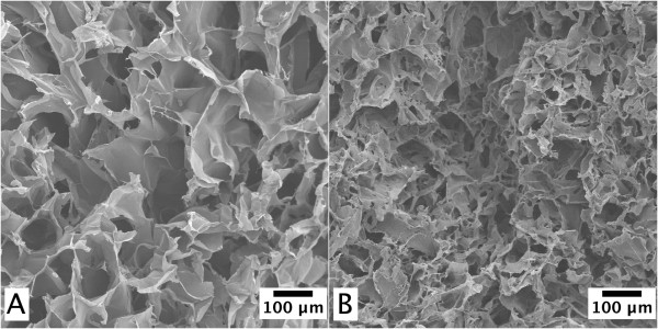
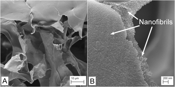
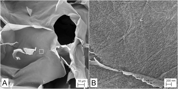
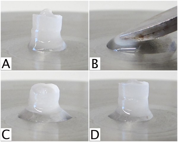
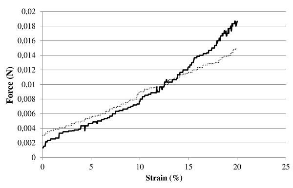
Cellulose nanofibrils were produced from P. radiata kraft pulp fibers. The nanofibrillation was facilitated by applying 2,2,6,6-tetramethylpiperidinyl-1-oxyl-mediated oxidation as pretreatment. The oxidized nanofibrils were cross-linked with polyethyleneimine and poly N-isopropylacrylamide-co-allylamine-co-methylenebisacrylamide particles and were frozen to form cryo-structured gels. Samples of the gels were critical-point dried, and the corresponding structures were assessed with scanning electron microscopy. It appears that the aldehyde groups in the oxidized nanofibrils are suitable reaction sites for cross-linking. The cryo-structured materials were spongy, elastic, and thus capable of regaining their shape after a given pressure was released, indicating a successful cross-linking. These novel types of gels are considered potential candidates in biomedical and biotechnological applications.
Figures

References
-
- Henriksson M, Berglund L. Structure and properties of cellulose nanocomposite films containing melamine formaldehyde. J Appl Polym Sci. 2007;106:2817–2824.
-
- Nakagaito AN, Yano H. Novel high-strength biocomposites based on microfibrillated cellulose having nano-order-unit web-like network structure. Appl Phys A. 2005;80:155–159.
-
- Bruce DM, Hobson RN, Farrent JW, Hepworth DG. High-performance composites from low-cost plant primary cell walls. Compos Part A. 2005;26:1486–1493.
-
- Malainine ME, Mahrouz M, Dufresne A. Thermoplastic nanocomposites based on cellulose microfibrils from Opuntia ficus-indica parenchyma cell. Compos Sci Technol. 2005;65:1520–1526.
LinkOut - more resources
Full Text Sources
Other Literature Sources
