

Identification of α(1,6)fucosylated proteins differentially expressed in human colorectal cancer
- PMID: 22152070
- PMCID: PMC3297542
- DOI: 10.1186/1471-2407-11-508
Identification of α(1,6)fucosylated proteins differentially expressed in human colorectal cancer
Abstract
Background: A universal hallmark of cancer cells is the change in their glycosylation phenotype. One of the most frequent alterations in the normal glycosylation pattern observed during carcinogenesis is the enhancement of α(1,6)linked fucose residues of glycoproteins, due to the up-regulation of the α(1,6)fucosyltransferase activity. Our previous results demonstrated the specific alteration of this enzyme activity and expression in colorectal cancer, suggesting its implication in tumour development and progression.
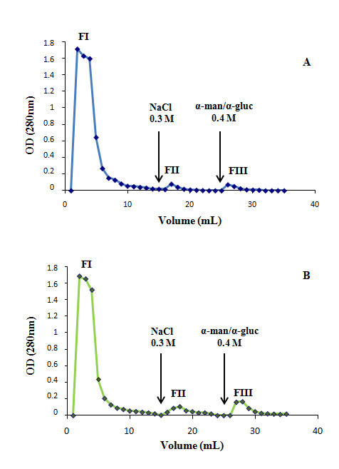
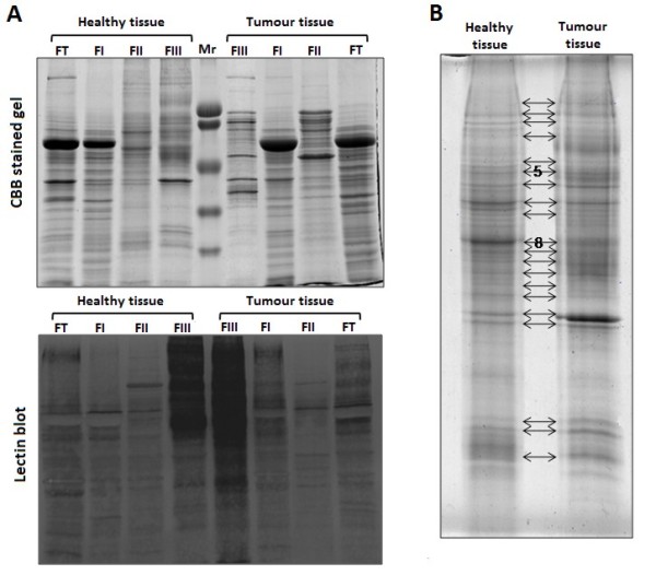
Methods: In the current work we combined a LCA-affinity chromatography with SDS-PAGE and mass spectrometry in order to identify α(1,6)fucosylated proteins differentially expressed in colorectal cancer. This strategy allowed the identification of a group of α(1,6)fucosylated proteins candidates to be involved in CRC malignancy.
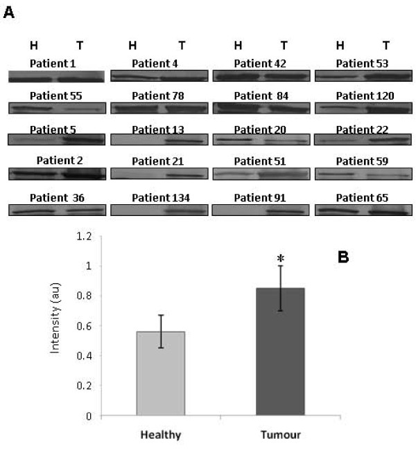
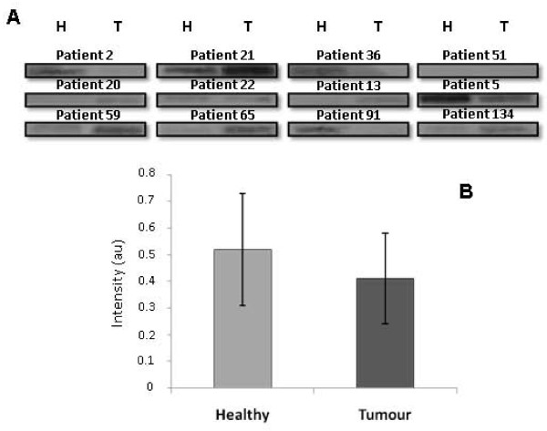
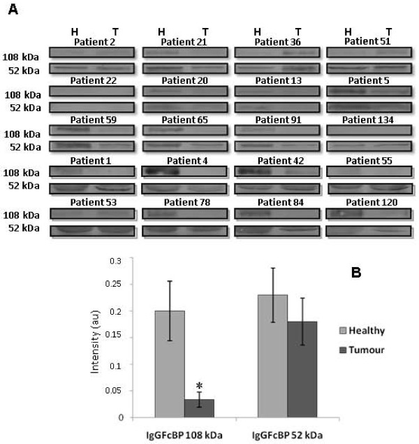
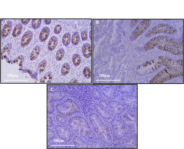
Results: The majority of the identified proteins take part in cell signaling and interaction processes as well as in modulation of the immunological response. Likewise, we confirmed the increased expression of GRP94 in colorectal cancer tissue and the significant down-regulation of the IgGFcBP expression in tumour cells.
Conclusion: All these results validate the importance of core-fucosylated proteins profile analysis to understand the mechanisms which promote cancer onset and progression and to discover new tumour markers or therapeutic targets.
Figures
Similar articles
-
Identification of O-Linked Glycoproteins Binding to the Lectin Helix pomatia Agglutinin as Markers of Metastatic Colorectal Cancer.PLoS One. 2015 Oct 23;10(10):e0138345. doi: 10.1371/journal.pone.0138345. eCollection 2015. PLoS One. 2015. PMID: 26495974 Free PMC article.
-
Identification of differentially-expressed proteins between early submucosal non-invasive and invasive colorectal cancer using 2D-DIGE and mass spectrometry.Int J Immunopathol Pharmacol. 2011 Oct-Dec;24(4):849-59. doi: 10.1177/039463201102400404. Int J Immunopathol Pharmacol. 2011. PMID: 22230392
-
Wnt-driven LARGE2 mediates laminin-adhesive O-glycosylation in human colonic epithelial cells and colorectal cancer.Cell Commun Signal. 2020 Jun 25;18(1):102. doi: 10.1186/s12964-020-00561-6. Cell Commun Signal. 2020. PMID: 32586342 Free PMC article.
-
Aberrant protein glycosylation in the colon adenoma-cancer sequence: Colorectal cancer mechanisms and clinical implications.Biochim Biophys Acta Mol Basis Dis. 2025 Aug;1871(6):167853. doi: 10.1016/j.bbadis.2025.167853. Epub 2025 Apr 17. Biochim Biophys Acta Mol Basis Dis. 2025. PMID: 40250777 Review.
-
Identifying N-Glycan Biomarkers in Colorectal Cancer by Mass Spectrometry.Acc Chem Res. 2016 Oct 18;49(10):2099-2106. doi: 10.1021/acs.accounts.6b00193. Epub 2016 Sep 21. Acc Chem Res. 2016. PMID: 27653471 Review.
Cited by
-
Recombinant Lectin from Tepary Bean (Phaseolus acutifolius) with Specific Recognition for Cancer-Associated Glycans: Production, Structural Characterization, and Target Identification.Biomolecules. 2020 Apr 23;10(4):654. doi: 10.3390/biom10040654. Biomolecules. 2020. PMID: 32340396 Free PMC article.
-
Glycan array analysis of Pholiota squarrosa lectin and other fucose-oriented lectins.Glycobiology. 2021 May 3;31(4):459-476. doi: 10.1093/glycob/cwaa093. Glycobiology. 2021. PMID: 33021632 Free PMC article.
-
Targeting deeper the human serum fucome by a liquid-phase multicolumn platform in combination with combinatorial peptide ligand libraries.J Chromatogr B Analyt Technol Biomed Life Sci. 2014 Mar 1;951-952:135-42. doi: 10.1016/j.jchromb.2014.01.037. Epub 2014 Jan 30. J Chromatogr B Analyt Technol Biomed Life Sci. 2014. PMID: 24556279 Free PMC article.
-
Inhibition of α(1,6)fucosyltransferase: Effects on Cell Proliferation, Migration, and Adhesion in an SW480/SW620 Syngeneic Colorectal Cancer Model.Int J Mol Sci. 2022 Jul 30;23(15):8463. doi: 10.3390/ijms23158463. Int J Mol Sci. 2022. PMID: 35955598 Free PMC article.
-
Mass spectrometry-based analysis of glycoproteins and its clinical applications in cancer biomarker discovery.Clin Proteomics. 2014 Apr 10;11(1):14. doi: 10.1186/1559-0275-11-14. Clin Proteomics. 2014. PMID: 24722010 Free PMC article.
References
-
- Hakomori S. Aberrant glycosylation in tumors and tumor-associated carbohydrate antigens. Adv Cancer Res. 1989;52:257–331. - PubMed
-
- Campion B, Leger D, Wieruszeski JM, Montreuil J, Spik G. Presence of fucosylated triantennary, tetraantennary and pentaantennary glycans in transferrin synthesized by the human hepatocarcinoma cell line Hep G2. Eur J Biochem. 1989;184(2):405–413. doi: 10.1111/j.1432-1033.1989.tb15032.x. - DOI - PubMed
-
- Uozumi N, Teshima T, Yamamoto T, Nishikawa A, Gao YE, Miyoshi E, Gao CX, Noda K, Islam KN, Ihara Y. et al.A fluorescent assay method for GDP-L-Fuc:N-acetyl-beta-D-glucosaminide alpha 1-6fucosyltransferase activity, involving high performance liquid chromatography. J Biochem. 1996;120(2):385–392. - PubMed
Publication types
MeSH terms
Substances
LinkOut - more resources
Full Text Sources
Medical
Research Materials
Miscellaneous
