Increased circulating leukocyte numbers and altered macrophage phenotype correlate with the altered immune response to brain injury in metallothionein (MT)-I/II null mutant mice
- PMID: 22152221
- PMCID: PMC3251619
- DOI: 10.1186/1742-2094-8-172
Increased circulating leukocyte numbers and altered macrophage phenotype correlate with the altered immune response to brain injury in metallothionein (MT)-I/II null mutant mice
Abstract
Background: Metallothionein-I and -II (MT-I/II) is produced by reactive astrocytes in the injured brain and has been shown to have neuroprotective effects. The neuroprotective effects of MT-I/II can be replicated in vitro which suggests that MT-I/II may act directly on injured neurons. However, MT-I/II is also known to modulate the immune system and inflammatory processes mediated by the immune system can exacerbate brain injury. The present study tests the hypothesis that MT-I/II may have an indirect neuroprotective action via modulation of the immune system.
Methods: Wild type and MT-I/II(-/-) mice were administered cryolesion brain injury and the progression of brain injury was compared by immunohistochemistry and quantitative reverse-transcriptase PCR. The levels of circulating leukocytes in the two strains were compared by flow cytometry and plasma cytokines were assayed by immunoassay.
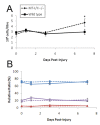
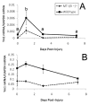
Results: Comparison of MT-I/II(-/-) mice with wild type controls following cryolesion brain injury revealed that the MT-I/II(-/-) mice only showed increased rates of neuron death after 7 days post-injury (DPI). This coincided with increases in numbers of T cells in the injury site, increased IL-2 levels in plasma and increased circulating leukocyte numbers in MT-I/II(-/-) mice which were only significant at 7 DPI relative to wild type mice. Examination of mRNA for the marker of alternatively activated macrophages, Ym1, revealed a decreased expression level in circulating monocytes and brain of MT-I/II(-/-) mice that was independent of brain injury.
Conclusions: These results contribute to the evidence that MT-I/II(-/-) mice have altered immune system function and provide a new hypothesis that this alteration is partly responsible for the differences observed in MT-I/II(-/-) mice after brain injury relative to wild type mice.
Figures


Similar articles
-
Strongly compromised inflammatory response to brain injury in interleukin-6-deficient mice.Glia. 1999 Feb 15;25(4):343-57. Glia. 1999. PMID: 10028917
-
Metallothionein (MT) -I and MT-II expression are induced and cause zinc sequestration in the liver after brain injury.PLoS One. 2012;7(2):e31185. doi: 10.1371/journal.pone.0031185. Epub 2012 Feb 17. PLoS One. 2012. PMID: 22363575 Free PMC article.
-
Metallothionein-1+2 protect the CNS after a focal brain injury.Exp Neurol. 2002 Jan;173(1):114-28. doi: 10.1006/exnr.2001.7772. Exp Neurol. 2002. PMID: 11771944
-
[Metallothionein-I/II in brain injury repair mechanism and its application in forensic medicine].Fa Yi Xue Za Zhi. 2013 Oct;29(5):365-7, 377. Fa Yi Xue Za Zhi. 2013. PMID: 24466778 Review. Chinese.
-
New insight into the molecular pathways of metallothionein-mediated neuroprotection and regeneration.J Neurochem. 2008 Jan;104(1):14-20. doi: 10.1111/j.1471-4159.2007.05026.x. Epub 2007 Nov 6. J Neurochem. 2008. PMID: 17986229 Review.
Cited by
-
Tumor cell cytoplasmic metallothionein expression associates with differential tumor immunogenicity and prognostic outcome in high-grade serous ovarian carcinoma.Front Oncol. 2023 Nov 8;13:1252700. doi: 10.3389/fonc.2023.1252700. eCollection 2023. Front Oncol. 2023. PMID: 38023247 Free PMC article.
-
The Role of Zinc and Zinc Homeostasis in Macrophage Function.J Immunol Res. 2018 Dec 6;2018:6872621. doi: 10.1155/2018/6872621. eCollection 2018. J Immunol Res. 2018. PMID: 30622979 Free PMC article. Review.
-
β1-Integrin Deletion From the Lens Activates Cellular Stress Responses Leading to Apoptosis and Fibrosis.Invest Ophthalmol Vis Sci. 2017 Aug 1;58(10):3896-3922. doi: 10.1167/iovs.17-21721. Invest Ophthalmol Vis Sci. 2017. PMID: 28763805 Free PMC article.
-
Vitamin D treatment during pregnancy prevents autism-related phenotypes in a mouse model of maternal immune activation.Mol Autism. 2017 Mar 7;8:9. doi: 10.1186/s13229-017-0125-0. eCollection 2017. Mol Autism. 2017. PMID: 28316773 Free PMC article.
-
Novel Anti-Neuroinflammatory Properties of a Thiosemicarbazone-Pyridylhydrazone Copper(II) Complex.Int J Mol Sci. 2022 Sep 14;23(18):10722. doi: 10.3390/ijms231810722. Int J Mol Sci. 2022. PMID: 36142627 Free PMC article.
References
Publication types
MeSH terms
Substances
LinkOut - more resources
Full Text Sources
Other Literature Sources
Molecular Biology Databases
Miscellaneous

