

3D Echo systematically underestimates right ventricular volumes compared to cardiovascular magnetic resonance in adult congenital heart disease patients with moderate or severe RV dilatation
- PMID: 22152255
- PMCID: PMC3283510
- DOI: 10.1186/1532-429X-13-78
3D Echo systematically underestimates right ventricular volumes compared to cardiovascular magnetic resonance in adult congenital heart disease patients with moderate or severe RV dilatation
Abstract
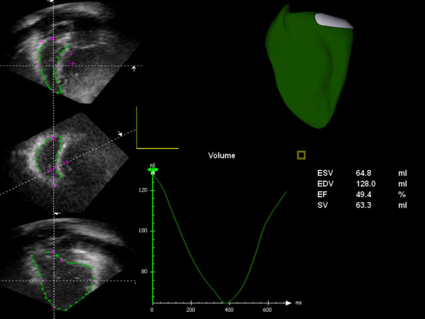
Background: Three dimensional echo is a relatively new technique which may offer a rapid alternative for the examination of the right heart. However its role in patients with non-standard ventricular size or anatomy is unclear. This study compared volumetric measurements of the right ventricle in 25 patients with adult congenital heart disease using both cardiovascular magnetic resonance (CMR) and three dimensional echocardiography.
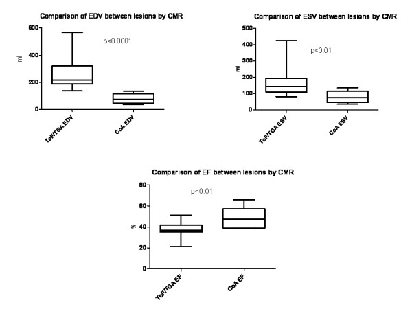
Methods: Patients were grouped by diagnosis into those expected to have normal or near-normal RV size (patients with repaired coarctation of the aorta) and patients expected to have moderate or worse RV enlargement (patients with repaired tetralogy of Fallot or transposition of the great arteries). Right ventricular end diastolic volume, end systolic volume and ejection fraction were compared using both methods with CMR regarded as the reference standard
Results: Bland-Altman analysis of the 25 patients demonstrated that for both RV EDV and RV ESV, there was a significant and systematic under-estimation of volume by 3D echo compared to CMR. This bias led to a mean underestimation of RV EDV by -34% (95%CI: -91% to + 23%). The degree of underestimation was more marked for RV ESV with a bias of -42% (95%CI: -117% to + 32%). There was also a tendency to overestimate RV EF by 3D echo with a bias of approximately 13% (95% CI -52% to +27%).
Conclusions: Statistically significant and clinically meaningful differences in volumetric measurements were observed between the two techniques. Three dimensional echocardiography does not appear ready for routine clinical use in RV assessment in congenital heart disease patients with more than mild RV dilatation at the current time.
Figures

Similar articles
-
Right Ventricular Assessment in Adult Congenital Heart Disease Patients with Right Ventricle-to-Pulmonary Artery Conduits.J Am Soc Echocardiogr. 2015 May;28(5):522-32. doi: 10.1016/j.echo.2014.11.016. Epub 2015 Jan 30. J Am Soc Echocardiogr. 2015. PMID: 25648672
-
Reproducibility of Cardiac Magnetic Resonance Imaging (CMRI)-Derived Right Ventricular Parameters in Repaired Tetralogy of Fallot (ToF).Heart Lung Circ. 2018 Mar;27(3):381-385. doi: 10.1016/j.hlc.2017.04.017. Epub 2017 Jun 3. Heart Lung Circ. 2018. PMID: 28662918
-
Can simple echocardiographic measures reduce the number of cardiac magnetic resonance imaging studies to diagnose right ventricular enlargement in congenital heart disease?J Am Soc Echocardiogr. 2012 May;25(5):518-23. doi: 10.1016/j.echo.2012.01.023. Epub 2012 Feb 23. J Am Soc Echocardiogr. 2012. PMID: 22365707
-
Review of the role of cardiovascular magnetic resonance in congenital heart disease, with a focus on right ventricle assessment.Arch Cardiovasc Dis. 2012 Nov;105(11):605-13. doi: 10.1016/j.acvd.2012.04.005. Epub 2012 Oct 4. Arch Cardiovasc Dis. 2012. PMID: 23177489 Review.
-
Assessment of right ventricular systolic function by echocardiography after surgical repair of congenital heart defects.Arch Cardiovasc Dis. 2016 Feb;109(2):113-9. doi: 10.1016/j.acvd.2015.11.002. Epub 2016 Jan 13. Arch Cardiovasc Dis. 2016. PMID: 26774976 Review.
Cited by
-
Echocardiographic assessment after surgical repair of tetralogy of fallot.Front Pediatr. 2015 Feb 2;3:3. doi: 10.3389/fped.2015.00003. eCollection 2015. Front Pediatr. 2015. PMID: 25699243 Free PMC article. Review.
-
SCMR Position Paper (2020) on clinical indications for cardiovascular magnetic resonance.J Cardiovasc Magn Reson. 2020 Nov 9;22(1):76. doi: 10.1186/s12968-020-00682-4. J Cardiovasc Magn Reson. 2020. PMID: 33161900 Free PMC article. Review.
-
Evaluation of right ventricular volume and ejection fraction by gated (18)F-FDG PET in patients with pulmonary hypertension: comparison with cardiac MRI and CT.J Nucl Cardiol. 2013 Apr;20(2):242-52. doi: 10.1007/s12350-013-9672-8. Epub 2013 Jan 26. J Nucl Cardiol. 2013. PMID: 23354658
-
Reply to Vanderpool et al.Am J Respir Crit Care Med. 2023 Apr 15;207(8):1102-1103. doi: 10.1164/rccm.202301-0100LE. Am J Respir Crit Care Med. 2023. PMID: 36689753 Free PMC article. No abstract available.
-
Cardiovascular Magnetic Resonance Imaging: A Prospective Modality in the Diagnosis and Prognostication of Heart Failure.Cureus. 2022 Apr 5;14(4):e23840. doi: 10.7759/cureus.23840. eCollection 2022 Apr. Cureus. 2022. PMID: 35530891 Free PMC article. Review.
References
-
- Gatzoulis MA, Balaji S, Webber SA, Siu SC, Hokanson JS, Poile C, Rosenthal M, Nakazawa M, Moller JH, Gillette PC, Webb GD, Redington AN. Risk factors for arrhythmia and sudden cardiac death late after repair of tetralogy of Fallot: A multicentre study. Lancet. 2000;356(9234):975–81. doi: 10.1016/S0140-6736(00)02714-8. - DOI - PubMed
-
- Greutmann M, Tobler D, Biaggi P, Mah ML, Crean A, Oechslin EN, Silversides CK. Echocardiography for assessment of right ventricular volumes revisited: A cardiac magnetic resonance comparison study in adults with repaired tetralogy of Fallot. J Am Soc Echocardiogr. 2010;23(9):905–11. doi: 10.1016/j.echo.2010.06.013. - DOI - PubMed
-
- Rudski LG, Lai WW, Afilalo J, Hua L, Handschumacher MD, Chandrasekaran K, Solomon SD, Louie EK, Schiller NB. Guidelines for the echocardiographic assessment of the right heart in adults: A report from the American Society of Echocardiography endorsed by the European Association of Echocardiography, a registered branch of the European Society of Cardiology, and the Canadian Society of Echocardiography. J Am Soc Echocardiogr. 2010;23(7):685–713. doi: 10.1016/j.echo.2010.05.010. - DOI - PubMed
-
- van der Zwaan HB, Helbing WA, McGhie JS, Geleijnse ML, Luijnenburg SE, Roos-Hesselink JW, Meijboom FJ. Clinical value of real-time three-dimensional echocardiography for right ventricular quantification in congenital heart disease: Validation with cardiac magnetic resonance imaging. J Am Soc Echocardiogr. 2010;23(2):134–40. doi: 10.1016/j.echo.2009.12.001. - DOI - PubMed
Publication types
MeSH terms
LinkOut - more resources
Full Text Sources
Medical
Molecular Biology Databases
