. 2011 Dec 7:8:173.
doi: 10.1186/1742-2094-8-173.

Adaptive Müller cell responses to microglial activation mediate neuroprotection and coordinate inflammation in the retina

Affiliations

Adaptive Müller cell responses to microglial activation mediate neuroprotection and coordinate inflammation in the retina

Minhua Wang et al. J Neuroinflammation. .

Abstract

Purpose: Microglia and Müller cells are prominent participants in retinal responses to injury and disease that shape eventual tissue adaptation or damage. This investigation examined how microglia and Müller cells interact with each other following initial microglial activation.

Methods: Mouse Müller cells were cultured alone, or co-cultured with activated or unactivated retinal microglia, and their morphological, molecular, and functional responses were evaluated. Müller cell-feedback signaling to microglia was studied using Müller cell-conditioned media. Corroborative in vivo analyses of retinal microglia-Müller cell interactions in the mouse retina were also performed.

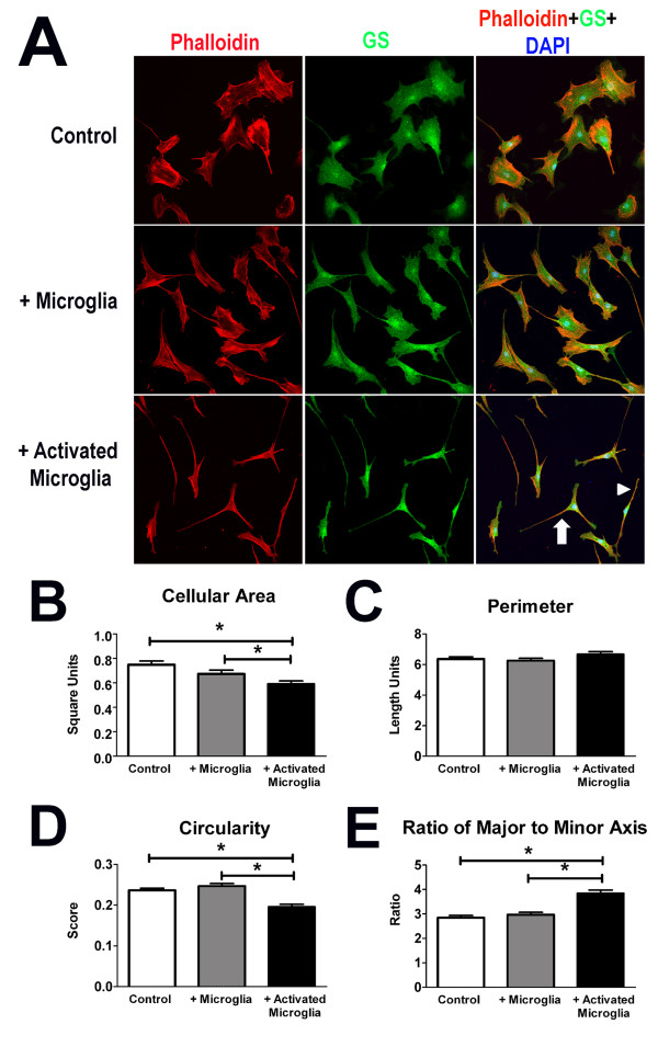
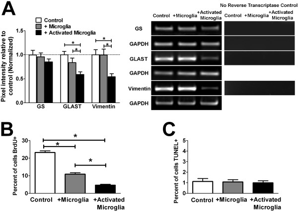
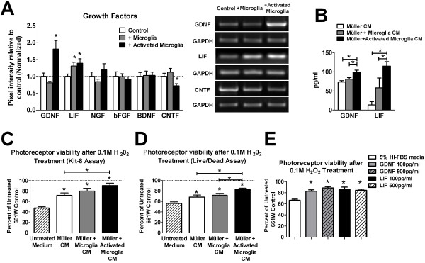
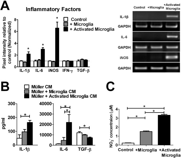
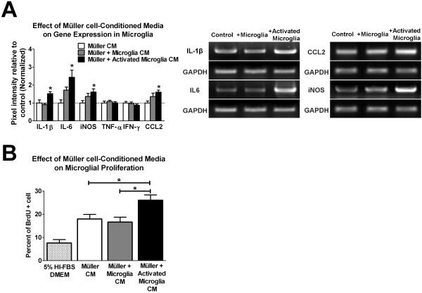
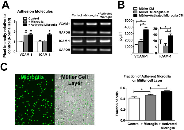
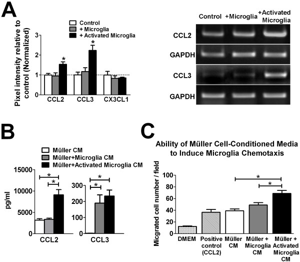
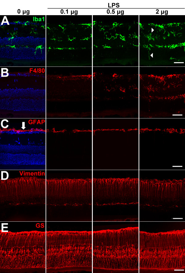
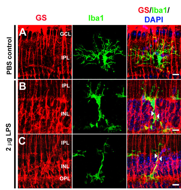
Results: Our results demonstrate that Müller cells exposed to activated microglia, relative to those cultured alone or with unactivated microglia, exhibit marked alterations in cell morphology and gene expression that differed from those seen in chronic gliosis. These Müller cells demonstrated in vitro (1) an upregulation of growth factors such as GDNF and LIF, and provide neuroprotection to photoreceptor cells, (2) increased pro-inflammatory factor production, which in turn increased microglial activation in a positive feedback loop, and (3) upregulated chemokine and adhesion protein expression, which allowed Müller cells to attract and adhere to microglia. In vivo activation of microglia by intravitreal injection of lipopolysaccharide (LPS) also induced increased Müller cell-microglia adhesion, indicating that activated microglia may translocate intraretinally in a radial direction using Müller cell processes as an adhesive scaffold.

Conclusion: Our findings demonstrate that activated microglia are able to influence Müller cells directly, and initiate a program of bidirectional microglia-Müller cell signaling that can mediate adaptive responses within the retina following injury. In the acute aftermath following initial microglia activation, Müller cell responses may serve to augment initial inflammatory responses across retinal lamina and to guide the intraretinal mobilization of migratory microglia using chemotactic cues and adhesive cell contacts. Understanding adaptive microglia-Müller cell interactions in injury responses can help discover therapeutic cellular targets for intervention in retinal disease.


Figures

Figure 1
Figure 1
Microglia-coculture induces alterations in cellular morphology in cultured Müller cells. (A) Morphological features of cultured mouse Müller cells were visualized by immunolabelling of the actin cytoskeleton (with phalloidin, red), cytoplasm (with glutamine synthetase (GS), green), and nuclei (with DAPI, blue). Representative examples of Müller cells cultured alone (top), co-cultured with mouse microglia (middle), and cultured with microglia previously activated with lipopolysaccharide (LPS, 1 μg/ml) (bottom), are shown. Müller cells cultured alone were similar to those co-cultured with unactivated microglia in having broad lamellipodia and a symmetrical cell shape (top and middle rows) while co-cultured with activated microglia (bottom row) had elongated spindle (arrowhead) or multipolar (arrow) morphologies. Quantitative shape analyses in terms of morphological parameters of cellular area (B), perimeter (C), circularity (D) and elongation factor (E) revealed significant decreases in cellular area and increases in cellular elongation, without changes in overall cellular perimeter (n = 348 to 363 cells per group, * indicates p values < 0.05 for comparisons, one-way ANOVA).
Figure 2
Figure 2
Influence of microglia on Müller cell gliosis, proliferation, and apoptosis. (A) The influence of microglia on Müller cell gliosis was assessed by evaluating mRNA expression of genes typically altered in gliosis by semi-quantitative RT-PCR: glutamine synthethase (GS), glutamate aspartate transporter (GLAST), and the intermediate filament, vimentin. Representative gel images for the PCR amplification of each mRNA species, with parallel controls from which reverse transcriptase is omitted from the amplification reaction (to confirm the absence of genomic DNA amplification), are shown (right). While the expression levels of GS in Müller cells were not statistically distinct between the different co-culture conditions, levels GLAST and vimentin, typically elevated in Müller cell gliosis, were significantly decreased following co-culture with activated microglia. These results demonstrate that changes induced by microglia co-culture differed from those associated with typical Müller cells gliosis. (B) Proliferating Müller cells in culture were marked by the incorporation of BrdU and the number of proliferating cells counted and expressed as a percentage of cells present. Co-culture with unactivated microglia induced a significant decrease in Müller cell proliferation, which was further decreased with co-culture with activated microglia. (C) Müller cells undergoing apoptosis in culture were marked with TUNEL-labeling. The percentage of apoptotic Müller cells was low and similar between all three co-culture conditions. (* indicates p < 0.05 for comparisons, one-way ANOVA with Tukey-Kramer multiple comparison test, n = 6-8 replicates from two independent experiments).
Figure 3
Figure 3
Müller cell expression of growth factors and neuroprotective function following microglial co-culture. (A) Semi-quantitative RT-PCR comparing mRNA levels of growth factors in Müller cells following microglial co-culture. Expression of growth factors GDNF and LIF were significantly elevated in Müller cells co-cultured with activated microglia. Expression levels of NGF, bFGF, and BDNF were similar between the three groups, while CNTF levels were slightly reduced. Representative gel images for genes whose expression were significantly changed following co-culture are shown (right). (B) ELISA quantification of protein levels of GDNF and LIF in the conditioned media of Müller cells following microglial co-culture. Relatively elevated levels of these growth factors were found in conditioned media from Müller cells co-cultured with activated microglia. (C, D) Neuroprotective function of Müller cells following microglial co-culture were evaluated by assessing the ability of conditioned media from co-cultured Müller cells to rescue photoreceptor cells (661W cells) from H202-induced oxidative cell death. Following Müller cell-microglia co-culture, microglia-containing cell inserts were removed, fresh medium was added to the resulting Müller cell cultures and left to condition for 24 hours. These conditioned media were added to 661W cells in the presence of 0.1 M H202. 661W cells exposed to 0.1 M H202 in regular unconditioned media served as controls. Cell viability of H202-exposed 661W cells were evaluated with a tetrazolium-based cell counting Kit-8 assay (in C) and a differential cell-staining (Live/Dead) assay (in D). Müller cell-media from all co-culture conditions exerted significant neuroprotective effect relative to unconditioned medium (marked by * over individual bars) but that from Müller cells co-cultured with activated microglia exerted a greater neuroprotective effect relative to Müller cells cultured without microglia. (E) Neuroprotective effect of exogenous GDNF and LIF on photoreceptor cells undergoing oxidative stress. GDNF and LIF (100 pg/ml and 500 pg/ml) were added to 661W cells in the presence of 0.1 M H202. 661W cells exposed to 0.1 M H202 in regular unconditioned media served as a control. Additions of GDNF and LIF were able to significantly increase 661W cell survival as evaluated with a cell counting Kit-8 assay. No significant dose-dependent effect was observed in the range of concentrations used. (* indicates p < 0.05 for comparisons, one-way ANOVA with Tukey-Kramer multiple comparison test, n = 6-8 replicates from two independent experiments).
Figure 4
Figure 4
Influence of microglia on Müller cell expression of inflammatory factors. (A) Semi-quantitative RT-PCR comparing mRNA levels of inflammatory factors in Müller cells cultured alone (white bars), or co-cultured with unactivated (gray bars) or LPS-activated (black bars) microglia. mRNA levels of inflammatory cytokines IL-1β and IL-6, as well as iNOS, were significantly elevated in Müller cell co-cultured with activated microglia, compared with those cultured alone or cultured with unactivated microglia. Expression levels of IFN-γ and TGF-β were similar between the three groups. Representative gel images for genes whose expression were significantly changed following co-culture are shown (right). (B) Protein levels of cytokines in the conditioned media of Müller cells 24 hours following co-culture were measured with ELISA. Inflammatory cytokines, IL-1β and IL-6, were both elevated, while levels of TGF-β were slightly decreased. (C) Nitrite concentrations in conditioned media following co-culture were elevated in Müller cells co-cultured with activated microglia, consistent with the increased expression for iNOS in Müller cells. (* indicates p < 0.05 for comparisons, one-way ANOVA with Tukey-Kramer multiple comparison test, n = 6-8 replicates from two independent experiments).
Figure 5
Figure 5
Müller cells, following co-culture with microglia, can induce reciprocal activation of retinal microglia. Following microglial co-culture, fresh media were added to Müller cells from each co-culture condition, left to condition for 24 hours, and then collected and tested for the ability to induce microglial activation. These conditioned media were added to fresh, unactivated cultured microglia for 24 hours. The ability of conditioned media to induce reciprocal microglial activation was assessed by measuring microglial mRNA expression of proinflammatory factors and microglial proliferation. (A) Conditioned media from Müller cells co-cultured with activated microglia were able to induce the highest levels of IL-1β, IL-6, iNOS, and CCL2 expression in microglia. Microglial expression levels of TNF-α and IFN-γ remained unchanged between three groups. Representative gel images for genes whose expression were significantly changed following co-culture are shown (right). (B) Microglia proliferation, as measured by BrdU incorporation, was significantly elevated in the conditioned media from Müller cells exposed to activated microglia, relative to other conditioned media. (* indicates p < 0.05 for comparisons, one-way ANOVA with Tukey-Kramer multiple comparison test, n = 6-8 replicates from two independent experiments).
Figure 6
Figure 6
Influence of microglia on Müller cell expression of adhesion molecules and adhesion properties. (A) Semi-quantitative RT-PCR comparing mRNA levels of adhesion molecules, VCAM-1 and ICAM-1, in Müller cells cultured alone (control, white bars), co-cultured with unactivated (gray bars) or activated (black bars) microglia. The mRNA levels of VCAM-1 were significantly elevated in Müller cells co-cultured with unactivated and activated microglia, compared with Müller cells cultured alone. ICAM-1 was significantly elevated only in Müller cells that were co-cultured with activated microglia. Representative gel images for genes whose expression were significantly changed following co-culture are shown (right). (B) Protein levels of adhesion molecules in Müller cell-conditioned media were measured with ELISA. Relatively elevated levels of VCAM-1 and ICAM-1 were found in conditioned media from Müller cells co-cultured with activated microglia. (C) The ability of Müller cells following co-culture to function as an adhesive substrate for microglia cells was assessed by a cell-adhesion assay. Unactivated microglia, labeled vitally with Calcein-AM (left panel, in green), were seeded onto Müller cells following 48-hr co-culture and allowed to adhere (right panel, Müller cells seen in bright-field). Non-adherent microglia were washed off and the remaining cells were counted and expressed as a fraction of total microglial cells added. Müller cells that had previously been co-cultured with activated microglia were found to induce the highest levels of adhesion, followed by Müller cells that had previously been co-cultured with unactivated microglia. (* indicates p < 0.05 for comparisons, one-way ANOVA with Tukey-Kramer multiple comparison test, n = 6-8 replicates from two independent experiments).
Figure 7
Figure 7
Müller cell expression of chemotactic cytokines following microglia co-culture. (A) Quantitative RT-PCR comparing mRNA levels of chemotactic cytokines in Müller cells cultured alone (control, white bars), or co-cultured with unactivated (gray bars) or activated microglia (black bars). The mRNA levels of CCL2 and CCL3 were significantly elevated in Müller cell co-cultured with activated microglia, compared with those cultured alone or cultured with unactivated microglia. Expression levels of CX3CL1 were unchanged between the three groups. Representative gel images for genes whose expression were significantly changed following co-culture are shown (right). (B) Protein levels of chemokines CCL2 and CCL3 in Müller cell-conditioned media were measured with ELISA. Relatively elevated levels of these chemokines were found in conditioned media from Müller cells co-cultured with activated microglia. (C) The ability of Müller cell-conditioned media to induce the chemotaxis of microglia was assayed using a modified Boydon chamber. Conditioned media from Müller cells exposed to activated microglia exerted the largest chemoattractive effect on microglia (black bar), followed by media from Müller cells co-cultured with unactivated microglia (gray bar), relative to media from Müller cells that were cultured alone (white bar). CCL2 (100 nM in DMEM medium) was used as a positive control for microglial chemotaxis. (* indicates p < 0.05 for all comparisons, one-way ANOVA with Tukey-Kramer multiple comparison test, n = 6-8 replicates from two independent experiments.)).
Figure 8
Figure 8
In vivo retinal microglia and Müller cell responses to retinal microglial activation induced by intravitreal injection of lipopolysaccharide (LPS). In vivo activation of retinal microglia was induced by intravitreal injection of different total amounts of LPS (0 μg, 0.1 μg, 0.5 μg, 2 μg) dissolved in 1 × PBS. Eyes injected with only 1 × PBS (0 μg LPS) served as controls (left column). Animals were sacrificed and enucleated 3 days after intravitreal injection and cryosections prepared from the globes. (A) Iba1 immunolabeling (green) of retinal sections show that following LPS injection, microglia exhibit a dose-dependent increase in cell density and Iba-1 immunopositivity, as well as an increase in the number of vertically oriented processes (arrowheads) as compared to PBS-injected controls. (B) F4/80 immunolabeling (red), a marker of microglial activation, demonstrates a dose-dependent increase in the density of immunoreactive microglial cells following LPS injection. (C) GFAP immunolabeling (red) located only in astrocytic processes (arrow) in the PBS-injected control, did not change in its localization following LPS injection, indicating that typical Müller cell gliosis, exemplified by increased GFAP expression, did not occur under these conditions. Vimentin (D) and glutamine synthetase (GS) (E) immunolabeling (red), located in Müller cell process, was present under all conditions and were not noticeably different in intensity. No marked changes in the morphology of Müller cells were noted. Scale bar = 50 μM.
Figure 9
Figure 9
Physical interaction of microglia-Müller cell processes following microglial activation in vivo. In vivo activation of retinal microglia was induced by intravitreal injection of lipopolysaccharide (LPS, 2 μg in 1 μl of 1 × PBS). Control eyes were injected with 1 μl of 1 × PBS alone. Retinal cryosections were prepared 3 days after injection and immunolabeled with glutamine synthetase (GS) (red) to mark Müller cell processes, Iba-1 (green) to mark microglia, and DAPI (blue) to mark retinal cell nuclei. (A) In PBS-injected control eyes, ramified processes of microglia in the inner retina are oriented predominantly in the horizontal plane of the retina and show minimal interaction or fasciculation with the vertically oriented, GS-positive, Müller cell processes. (B, C) In LPS-injected eyes, microglia in the inner retina demonstrate a more vertical orientation of their processes compared to controls. Microglial processes were observed to be juxtaposed in close physical association with parallel Müller cell processes. Close fasciculation between the vertical processes of both cell types can be observed (arrowheads), suggesting cellular adhesion and physical interaction between Müller cell-microglia processes. In examples in which vertically oriented microglia appear to be migrating in the radial direction, Müller cell processes appear to be acting as a adhesive scaffold that guide microglia orientation and translocation. Scale bar = 10 μM.

Similar articles

Cited by

References

    1. Buffo A, Rolando C, Ceruti S. Astrocytes in the damaged brain: molecular and cellular insights into their reactive response and healing potential. Biochem Pharmacol. 2010;79:77–89. doi: 10.1016/j.bcp.2009.09.014. - DOI - PubMed
    1. Ransohoff RM, Cardona AE. The myeloid cells of the central nervous system parenchyma. Nature. 2011;468:253–262. - PubMed
    1. Lotan M, Schwartz M. Cross talk between the immune system and the nervous system in response to injury: implications for regeneration. FASEB J. 1994;8:1026–1033. - PubMed
    1. Zhang D, Hu X, Qian L, O'Callaghan JP, Hong JS. Astrogliosis in CNS pathologies: is there a role for microglia? Mol Neurobiol. 2010;41:232–241. doi: 10.1007/s12035-010-8098-4. - DOI - PMC - PubMed
    1. Liu W, Tang Y, Feng J. Cross talk between activation of microglia and astrocytes in pathological conditions in the central nervous system. Life Sci. 2011;89:141–146. doi: 10.1016/j.lfs.2011.05.011. - DOI - PubMed

Substances