. 2011 Dec 7;14(6):747-57.
doi: 10.1016/j.cmet.2011.11.006.

TGR5 activation inhibits atherosclerosis by reducing macrophage inflammation and lipid loading

Affiliations

TGR5 activation inhibits atherosclerosis by reducing macrophage inflammation and lipid loading

Thijs W H Pols et al. Cell Metab. .

Abstract

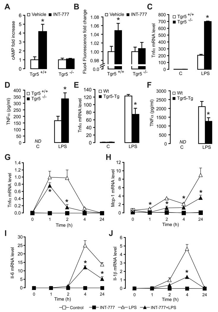
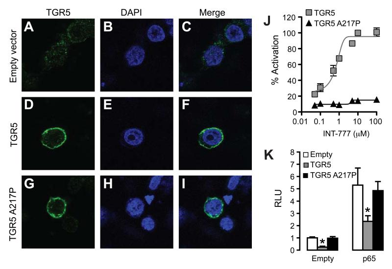
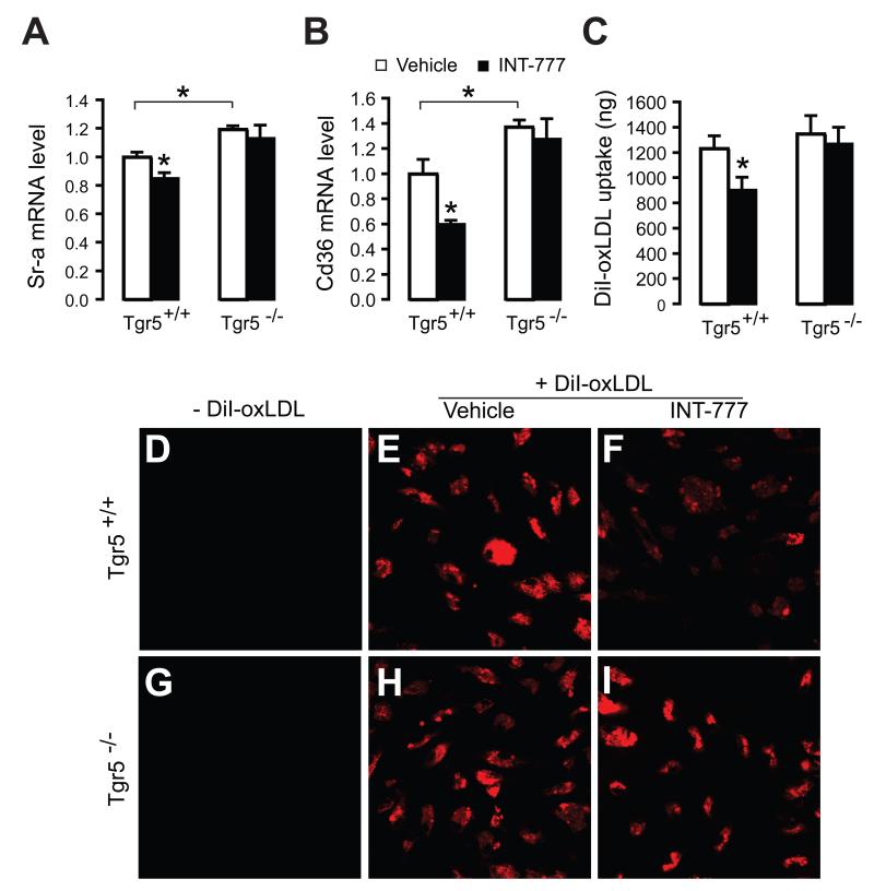
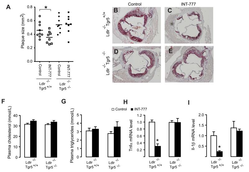
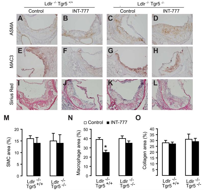
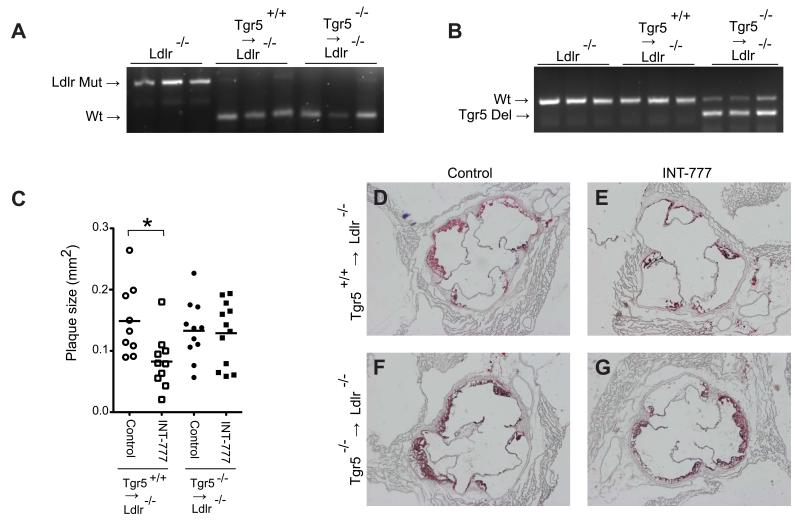
The G protein-coupled receptor TGR5 has been identified as an important component of the bile acid signaling network, and its activation has been linked to enhanced energy expenditure and improved glycemic control. Here, we demonstrate that activation of TGR5 in macrophages by 6α-ethyl-23(S)-methylcholic acid (6-EMCA, INT-777), a semisynthetic BA, inhibits proinflammatory cytokine production, an effect mediated by TGR5-induced cAMP signaling and subsequent NF-κB inhibition. TGR5 activation attenuated atherosclerosis in Ldlr(-/-)Tgr5(+/+) mice but not in Ldlr(-/-)Tgr5(-/-) double-knockout mice. The inhibition of lesion formation was associated with decreased intraplaque inflammation and less plaque macrophage content. Furthermore, Ldlr(-/-) animals transplanted with Tgr5(-/-) bone marrow did not show an inhibition of atherosclerosis by INT-777, further establishing an important role of leukocytes in INT-777-mediated inhibition of vascular lesion formation. Taken together, these data attribute a significant immune modulating function to TGR5 activation in the prevention of atherosclerosis, an important facet of the metabolic syndrome.


Figures

Figure 1
Figure 1. The TGR5 agonist INT-777 inhibits macrophage inflammation
(A) cAMP induction in primary macrophages isolated from Tgr5−/− and Tgr5+/+ mice measured 1 hr after stimulation with vehicle (white bars) or 3 μM INT-777 (black bars); (n=3). (B) Intracellular calcium flux in primary macrophages isolated from Tgr5−/− and Tgr5+/+ mice measured 30 sec after addition of 3 μM INT-777 (n=3). (C and D) mRNA expression (C) and protein secretion (D) of TNFα in primary macrophages isolated from Tgr5+/+ (white bars) or Tgr5−/− mice (black bars) in response to stimulation with 100 ng/ml LPS for 6 hrs (n=3). (E and F) mRNA level (E) and protein level (F) of TNFα in macrophages isolated from Tgr5-Tg mice (black bars) and wildtype mice (white bars) stimulated with 100 ng/ml LPS for 6 hrs in combination with treatment of 30 μM INT-777. (G-J) Tnfα (G), Mcp-1 (H), Il-6 (I), and Il-1β (J) cytokine mRNA in response to 100 ng/ml LPS (triangles) or not stimulated (squares) treated with 30 μM INT-777 (black) or control-treated (white) in RAW264.7 macrophages (n=3). All conditions are present at all timepoints. Results represent the mean ± SEM. * Statistically significant, P<0.05.
Figure 2
Figure 2. TGR5 activation inhibits NF-κB activation via cAMP signaling
(A) Western blot of C-Jun, phosphorylated c-Jun (P-C-Jun) with tubulin as loading control, and NF-κB p65 western blot of nuclear extract with PARP-1 as loading control of RAW264.7 macrophages treated with 100 ng/ml LPS for 3 hrs in combination with 100 μM SQ22536 and 30 μM INT-777 (n=3). (B) Quantification of western blot band intensity of p65 corrected for the intensity of PARP-1 using image analysis software. (C) Western blot of phosphorylated IκBα, total IκBα, and tubulin as loading control of lysate of RAW264.7 macrophages treated with 100 ng/ml LPS for 1 hr in combination with 30 μM INT-777 (n=3). (D) Quantification of western blot band intensity of IκBα corrected for the intensity of tubulin using image analysis software. (E) NF-κB-p65 binding activity to its DNA response element after 3 hours LPS stimulation. (F-H) LPS-induced (6 hrs) NF-κB transcriptional activity in RAW264.7 macrophages electroporated with the NF-κB reporter plasmid in combination with electroporation of TGR5 (F) or shTGR5 (G) in the presence of 30 μM INT-777 (black bars) or vehicle (white bars) (n=3) (H) LPS-induced NF-κB transcriptional activity in RAW264.7 macrophages electroporated with the NF-κB reporter plasmid in combination with 30 μM INT-777 treatment or vehicle in the presence of 100 μM SQ22536 (grey bars), 20 μM 2′, 5′-dideoxyadenosine (black bars) or control conditions (white bars) (n=3). Results represent the mean ± SEM. * Statistically significant, P<0.05.
Figure 3
Figure 3. The TGR5 mutant TGR5-A217P, defective in inducing cAMP signaling, fails to inhibit NF-κB activity
(A-I) Confocal images of empty vector (A-C), mouse TGR5 (D and F), and TGR5-A217P mutant (G-I) -transfected CHO cells stained with DAPI (B, E, H), stained for TGR5 (A, D, G) or shown as merged images (C, F, I). (J) CREB transcriptional activity in CHO cells transfected with both a CRE reporter and TGR5 wildtype (grey squares) or TGR5-A217P mutant (black triangles) in response to INT-777 (n=3). (K) NF-κB transcriptional activity in CHO cells transfected with the NF-κB reporter plasmid in combination with empty vector (white bars), TGR5 (grey bars) and TGR5-A217P (black bars) with or without NF-κB p65 co-transfection (n=3). Results represent the mean ± SEM. * Statistically significant, P<0.05.
Figure 4
Figure 4. TGR5 inhibits oxidized LDL uptake
(A and B) Sr-a (A) and Cd36 (B) mRNA expression in macrophages isolated from Tgr5+/+ and Tgr5−/− mice in response to INT-777 treatment (black bars) or control-treated (white bars; n=3). (C) Fluorescence of DiI-labeled oxidized LDL extracted from macrophages isolated from Tgr5+/+ and Tgr5−/− mice in response to INT-777 (black bars) or control conditions (white bars; n=3) (D-I) Confocal fluorescent images of macrophages from Tgr5+/+ (D, E, F) and Tgr5−/− (G, H, I) mice treated with DiI-labeled oxidized LDL (E, F, H, I) in combination with INT-777 treatment (F, I). Results represent the mean ± SEM. * Statistically significant, P<0.05.
Figure 5
Figure 5. TGR5 activation inhibits atherosclerosis
(A) Plaque size in the aortic root of Ldlr−/−Tgr5+/+ (white symbols) and Ldlr−/−Tgr5−/− animals (black symbols) treated with INT-777 (squares) or control-treated (circles) (n=8-9). (B-E) Oil-red-O staining of atherosclerotic lesions in the aortic root of Ldlr−/−Tgr5+/+ (B and C) or Ldlr−/−Tgr5−/− animals (D and E) treated with INT-777 (C and E) or control-treated (B and D). (F and G) Plasma cholesterol (F) and triglycerides (G) of Ldlr−/−Tgr5+/+ and Ldlr−/−Tgr5−/− animals treated with INT-777 (black bars) or control-treated (white bars; n=8-9). (H-I) mRNA levels of (H) Tnfα and (I) Il-1β in aortic root lesions captured by laser capture micro-dissection of Ldlr−/−Tgr5+/+ and Ldlr−/−Tgr5−/− animals treated with INT-777 (black bars) or control-treated (white bars; n=3). Results represent the mean ± SEM. * Statistically significant, P<0.05.
Figure 6
Figure 6. TGR5 modulates plaque macrophage content
(A-D) ASMA staining, (E-H) MAC3 staining (I-L) Sirius red staining to detect smooth muscle cells, macrophages and collagen, respectively, in aortic root lesions of Ldlr−/−Tgr5−/− and Ldlr−/−Tgr5+/+ animals treated with or without INT-777. (M-O) Quantification of ASMA (M), MAC3 (N) and Sirius red (O) staining area using image analysis software of aortic root lesions of Ldlr−/−Tgr5−/− and Ldlr−/−Tgr5+/+ animals treated with (black bars) or without INT-777 (white bars; n=8-9). Results represent the mean ± SEM. * Statistically significant, P<0.05.
Figure 7
Figure 7. INT-777 inhibits atherosclerosis through activation of TGR5 in leukocytes
(A and B) PCR products showing the genotype of the Ldlr (A) and Tgr5 (B) locus in genomic DNA isolated from circulating white blood cells of Ldlr−/− animals as well as Ldlr−/− animals transplanted with Tgr5+/+ and Tgr5−/− bone marrow. (C) Plaque size in the aortic root of Ldlr−/− animals transplanted with Tgr5+/+ (white symbols) or Tgr5−/− bone marrow (black symbols) treated with INT-777 (squares) or control-treated (circles) (n=9-12). (D-G) Oil-red-O staining of atherosclerotic lesions in the aortic root of Ldlr−/− animals carrying Tgr5+/+ (D and E) or Tgr5−/− bone marrow (F and G) treated with INT-777 (E and G) or control-treated (D and F). * Statistically significant, P<0.05.

Comment in

References

    1. Arakawa M, Mita T, Azuma K, Ebato C, Goto H, Nomiyama T, Fujitani Y, Hirose T, Kawamori R, Watada H. Inhibition of monocyte adhesion to endothelial cells and attenuation of atherosclerotic lesion by a glucagon-like peptide-1 receptor agonist, exendin-4. Diabetes. 2010;59:1030–1037. - PMC - PubMed
    1. Evans KA, Budzik BW, Ross SA, Wisnoski DD, Jin J, Rivero RA, Vimal M, Szewczyk GR, Jayawickreme C, Moncol DL, et al. Discovery of 3-aryl-4- isoxazolecarboxamides as TGR5 receptor agonists. J Med Chem. 2009;52:7962–7965. - PubMed
    1. Febbraio M, Podrez EA, Smith JD, Hajjar DP, Hazen SL, Hoff HF, Sharma K, Silverstein RL. Targeted disruption of the class B scavenger receptor CD36 protects against atherosclerotic lesion development in mice. J Clin Invest. 2000;105:1049–1056. - PMC - PubMed
    1. Ferreira V, van Dijk KW, Groen AK, Vos RM, van der Kaa J, Gijbels MJ, Havekes LM, Pannekoek H. Macrophage-specific inhibition of NF-kappaB activation reduces foam-cell formation. Atherosclerosis. 2007;192:283–290. - PubMed
    1. Genet C, Strehle A, Schmidt C, Boudjelal G, Lobstein A, Schoonjans K, Souchet M, Auwerx J, Saladin R, Wagner A. Structure-activity relationship study of betulinic acid, a novel and selective TGR5 agonist, and its synthetic derivatives: potential impact in diabetes. J Med Chem. 2010;53:178–190. - PubMed

Publication types

MeSH terms