

Histological damage and inflammatory response elicited by Monobothrium wageneri (Cestoda) in the intestine of Tinca tinca (Cyprinidae)
- PMID: 22152408
- PMCID: PMC3261215
- DOI: 10.1186/1756-3305-4-225
Histological damage and inflammatory response elicited by Monobothrium wageneri (Cestoda) in the intestine of Tinca tinca (Cyprinidae)
Abstract
Background: Among the European cyprinids, tench, Tinca tinca (L.), and the pathological effects their cestodes may effect, have received very little or no attention. Most literature relating to Monobothrium wageneri Nybelin, 1922, a common intestinal cestode of tench, for example, has focused on aspects of its morphology rather than on aspects of the host-parasite interaction.
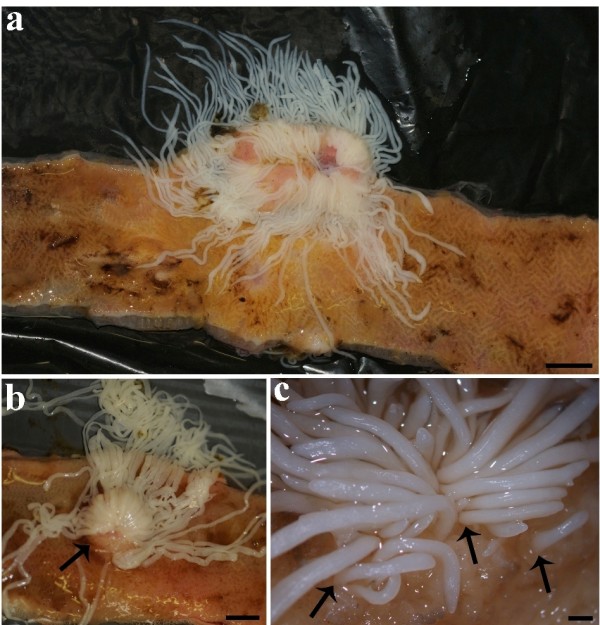
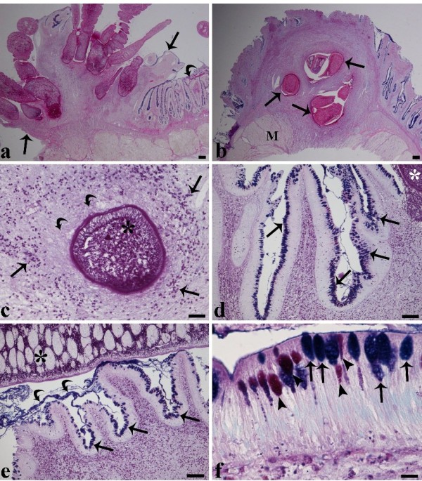
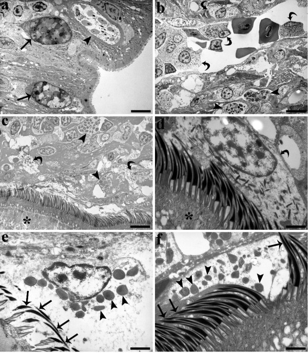
Results: Immunopathological and ultrastructural studies were conducted on the intestines of 28 tench, collected from Lake Piediluco, of which 16 specimens harboured tight clusters of numerous M. wageneri attached to the intestinal wall. The infection was associated with the degeneration of the mucosal layer and the formation of raised inflammatory swelling surrounding the worms. At the site of infection, the number of granulocytes in the intestine of T. tinca was significantly higher than the number determined 1 cm away from the site of infection or the number found in uninfected fish. Using transmission electron microscopy, mast cells and neutrophils were frequently observed in close proximity to, and inside, the intestinal capillaries; often these cells were in contact with the cestode tegument. At the host-parasite interface, no secretion from the parasite's tegument was observed. Intense degranulation of the mast cells was seen within the submucosa and lamina muscularis, most noticeably at sites close to the tegument of the scolex. In some instances, rodlet cells were encountered in the submucosa. In histological sections, hyperplasia of the mucous cells, notably those giving an alcian blue positive reaction, were evident in the intestinal tissues close to the swelling surrounding the worms. Enhanced mucus secretion was recorded in the intestines of infected tench.
Conclusions: The pathological changes and the inflammatory cellular response induced by the caryophyllidean monozoic tapeworm M. wageneri within the intestinal tract of an Italian population of wild tench is reported for the first time.
Figures
Similar articles
-
Innate immune defence mechanisms of tench, Tinca tinca (L.), naturally infected with the tapeworm Monobothrium wageneri.Parasite Immunol. 2012 Nov;34(11):511-9. doi: 10.1111/j.1365-3024.2012.01373.x. Parasite Immunol. 2012. PMID: 22709447
-
Histopathological and ultrastructural studies of the tapeworm Monobothrium wageneri (Caryophyllidea) in the intestinal tract of tench Tinca tinca.Dis Aquat Organ. 2011 Dec 6;97(2):143-54. doi: 10.3354/dao02406. Dis Aquat Organ. 2011. PMID: 22303631
-
New circumscription of freshwater fish parasites Monobothrium diesing, 1863 and Promonobothrium mackiewicz, 1968 (Cestoda: Caryophyllidea) using morphological and molecular evidence.J Parasitol. 2015 Feb;101(1):29-36. doi: 10.1645/14-610.1. Epub 2014 Sep 26. J Parasitol. 2015. PMID: 25259406
-
Heavy metal concentrations in adult acanthocephalans and cestodes compared to their fish hosts and to established free-living bioindicators.Parassitologia. 1997 Sep;39(3):213-8. Parassitologia. 1997. PMID: 9802069 Review.
-
Update on the distribution of the co-invasive Schyzocotyle acheilognathi (= Bothriocephalus acheilognathi), the Asian fish tapeworm, in freshwater fishes of Mexico.J Helminthol. 2018 May;92(3):279-290. doi: 10.1017/S0022149X17000438. Epub 2017 May 22. J Helminthol. 2018. PMID: 28528580 Review.
Cited by
-
Nematode infection in liver of the fish Gymnotus inaequilabiatus (Gymnotiformes: Gymnotidae) from the Pantanal Region in Brazil: pathobiology and inflammatory response.Parasit Vectors. 2016 Aug 30;9(1):473. doi: 10.1186/s13071-016-1772-2. Parasit Vectors. 2016. PMID: 27576434 Free PMC article.
-
New cell motility model observed in parasitic cnidarian Sphaerospora molnari (Myxozoa:Myxosporea) blood stages in fish.Sci Rep. 2016 Dec 16;6:39093. doi: 10.1038/srep39093. Sci Rep. 2016. PMID: 27982057 Free PMC article.
-
Enteric neuromodulators and mucus discharge in a fish infected with the intestinal helminth Pomphorhynchus laevis.Parasit Vectors. 2015 Jul 8;8:359. doi: 10.1186/s13071-015-0970-7. Parasit Vectors. 2015. PMID: 26152567 Free PMC article.
-
Histopathology and the inflammatory response of European perch, Perca fluviatilis muscle infected with Eustrongylides sp. (Nematoda).Parasit Vectors. 2015 Apr 15;8:227. doi: 10.1186/s13071-015-0838-x. Parasit Vectors. 2015. PMID: 25889096 Free PMC article.
-
Infection of endemic chub Squalius tenellus with the intestinal tapeworm Caryophyllaeus brachycollis (Cestoda): histopathology and ultrastructural surveys.Parasitology. 2024 Feb;151(2):157-167. doi: 10.1017/S0031182023001233. Epub 2023 Dec 1. Parasitology. 2024. PMID: 38193283 Free PMC article.
References
-
- Gibson DI. Monobothrium wageneri: another imported tapeworm established in wild British freshwater fishes? J Fish Biol. 1993;43:281–285. doi: 10.1111/j.1095-8649.1993.tb00428.x. - DOI
-
- Oros M, Scholz T, Hanzelová V, Mackiewicz JS. Scolex morphology of monozoic cestodes (Caryophyllidea) from the Palaearctic Region: a useful tool for species identification. Folia Parasitol. 2010;57:37–46. - PubMed
-
- Ringø E, Myklebust R, Mayhew TM, Olsen RE. Bacterial translocation and pathogenesis in the digestive tract of larvae and fry. Aquaculture. 2007;268:251–264. doi: 10.1016/j.aquaculture.2007.04.047. - DOI
Publication types
MeSH terms
LinkOut - more resources
Full Text Sources
