

Decreased CXCR1 and CXCR2 expression on neutrophils in anti-neutrophil cytoplasmic autoantibody-associated vasculitides potentially increases neutrophil adhesion and impairs migration
- PMID: 22152684
- PMCID: PMC3334654
- DOI: 10.1186/ar3534
Decreased CXCR1 and CXCR2 expression on neutrophils in anti-neutrophil cytoplasmic autoantibody-associated vasculitides potentially increases neutrophil adhesion and impairs migration
Abstract
Introduction: In anti-neutrophil cytoplasmic autoantibody (ANCA)-associated vasculitides (AAV), persistent inflammation within the vessel wall suggests perturbed neutrophil trafficking leading to accumulation of activated neutrophils in the microvascular compartment. CXCR1 and CXCR2, being major chemokine receptors on neutrophils, are largely responsible for neutrophil recruitment. We speculate that down-regulated expression of CXCR1/2 retains neutrophils within the vessel wall and, consequently, leads to vessel damage.
Methods: Membrane expression of CXCR1/2 on neutrophils was assessed by flow cytometry. Serum levels of interleukin-8 (IL-8), tumor necrosis factor alpha (TNF-α), angiopoietin 1 and angiopoietin 2 from quiescent and active AAV patients and healthy controls (HC) were quantified by ELISA. Adhesion and transendothelial migration of isolated neutrophils were analyzed using adhesion assays and Transwell systems, respectively.
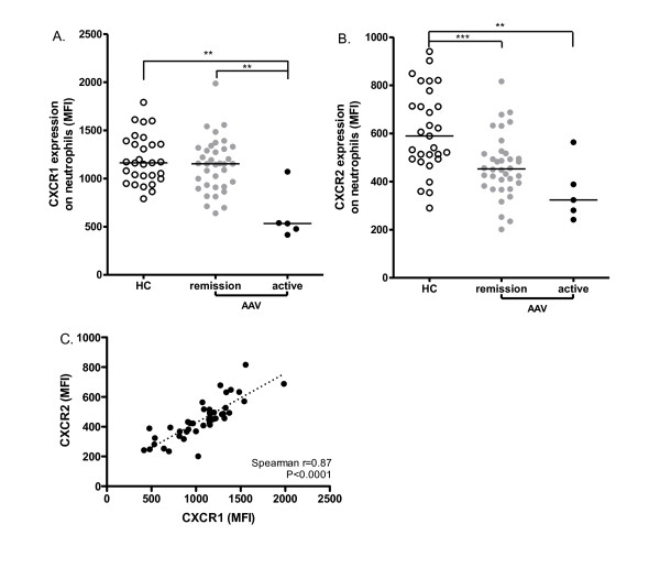
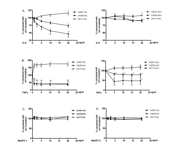
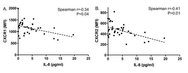
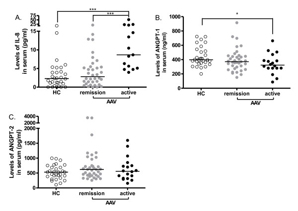
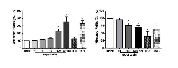
Results: Expression of CXCR1 and CXCR2 on neutrophils was significantly decreased in AAV patients compared to HC. Levels of IL-8, which, as TNFα, dose-dependently down-regulated CXCR1 and CXCR2 expression on neutrophils in vitro, were significantly increased in the serum of patients with active AAV and correlated negatively with CXCR1/CXCR2 expression on neutrophils, even in quiescent patients. Blocking CXCR1 and CXCR2 with repertaxin increased neutrophil adhesion and inhibited migration through a glomerular endothelial cell layer.
Conclusions: Expression of CXCR1 and CXCR2 is decreased in AAV, potentially induced by circulating proinflammatory cytokines such as IL-8. Down-regulation of these chemokine receptors could increase neutrophil adhesion and impair its migration through the glomerular endothelium, contributing to neutrophil accumulation and, in concert with ANCA, persistent inflammation within the vessel wall.
Figures
Similar articles
-
Elucidation of Distinct Roles of Guinea Pig CXCR1 and CXCR2 in Neutrophil Migration toward IL-8 and GROα by Specific Antibodies.Biol Pharm Bull. 2017;40(5):729-732. doi: 10.1248/bpb.b16-00918. Biol Pharm Bull. 2017. PMID: 28458362
-
ANCA induces beta2 integrin and CXC chemokine-dependent neutrophil-endothelial cell interactions that mimic those of highly cytokine-activated endothelium.J Leukoc Biol. 2005 Jan;77(1):33-43. doi: 10.1189/jlb.0104054. Epub 2004 Sep 30. J Leukoc Biol. 2005. PMID: 15459232
-
The chemokine receptors CXCR1/CXCR2 modulate antigen-induced arthritis by regulating adhesion of neutrophils to the synovial microvasculature.Arthritis Rheum. 2008 Aug;58(8):2329-37. doi: 10.1002/art.23622. Arthritis Rheum. 2008. PMID: 18668539
-
Combined anti CXC receptors 1 and 2 therapy is a promising anti-inflammatory treatment for respiratory diseases by reducing neutrophil migration and activation.Pulm Pharmacol Ther. 2015 Oct;34:37-45. doi: 10.1016/j.pupt.2015.08.002. Epub 2015 Aug 10. Pulm Pharmacol Ther. 2015. PMID: 26271598 Review.
-
A narrative review of chemokine receptors CXCR1 and CXCR2 and their role in acute respiratory distress syndrome.Eur Respir Rev. 2024 Jul 24;33(173):230172. doi: 10.1183/16000617.0172-2023. Print 2024 Jul. Eur Respir Rev. 2024. PMID: 39048127 Free PMC article. Review.
Cited by
-
Corticosteroid use and increased CXCR2 levels on leukocytes are associated with lumacaftor/ivacaftor discontinuation in cystic fibrosis patients homozygous for the F508del CFTR mutation.PLoS One. 2018 Dec 12;13(12):e0209026. doi: 10.1371/journal.pone.0209026. eCollection 2018. PLoS One. 2018. PMID: 30540818 Free PMC article.
-
Microphysiological Systems for Studying Cellular Crosstalk During the Neutrophil Response to Infection.Front Immunol. 2021 Apr 27;12:661537. doi: 10.3389/fimmu.2021.661537. eCollection 2021. Front Immunol. 2021. PMID: 33986752 Free PMC article. Review.
-
IL-8 Triggers Neutrophil Extracellular Trap Formation Through an Nicotinamide Adenine Dinucleotide Phosphate Oxidase- and Mitogen-Activated Protein Kinase Pathway-Dependent Mechanism in Uveitis.Invest Ophthalmol Vis Sci. 2023 Oct 3;64(13):19. doi: 10.1167/iovs.64.13.19. Invest Ophthalmol Vis Sci. 2023. PMID: 37824136 Free PMC article.
-
Factors regulating subcutaneous adipose tissue storage, fibrosis, and inflammation may underlie low fatty acid mobilization in insulin-sensitive obese adults.Am J Physiol Endocrinol Metab. 2017 Oct 1;313(4):E429-E439. doi: 10.1152/ajpendo.00084.2017. Epub 2017 Jul 5. Am J Physiol Endocrinol Metab. 2017. PMID: 28679624 Free PMC article.
-
The interaction between C5a and sphingosine-1-phosphate in neutrophils for antineutrophil cytoplasmic antibody mediated activation.Arthritis Res Ther. 2014 Jul 7;16(4):R142. doi: 10.1186/ar4604. Arthritis Res Ther. 2014. PMID: 25000985 Free PMC article.
References
MeSH terms
Substances
Grants and funding
LinkOut - more resources
Full Text Sources
Medical
