. 2011 Dec 13;58(25):2683-91.
doi: 10.1016/j.jacc.2011.09.033.

Beta3-adrenoreceptor stimulation ameliorates myocardial ischemia-reperfusion injury via endothelial nitric oxide synthase and neuronal nitric oxide synthase activation

Affiliations

Beta3-adrenoreceptor stimulation ameliorates myocardial ischemia-reperfusion injury via endothelial nitric oxide synthase and neuronal nitric oxide synthase activation

Juan P Aragón et al. J Am Coll Cardiol. .

Abstract

Objectives: This paper examined whether nebivolol protects the heart via nitric oxide (NO) synthase and NO-dependent signaling in an in vivo model of acute myocardial infarction.

Background: Beta(3)-adrenergic receptor (AR) activation promotes endothelial nitric oxide synthase (eNOS) activity and NO bioavailability. We hypothesized that specific beta(3)-AR agonists would attenuate myocardial ischemia-reperfusion (MI/R) injury via eNOS activation and increased NO bioavailability.

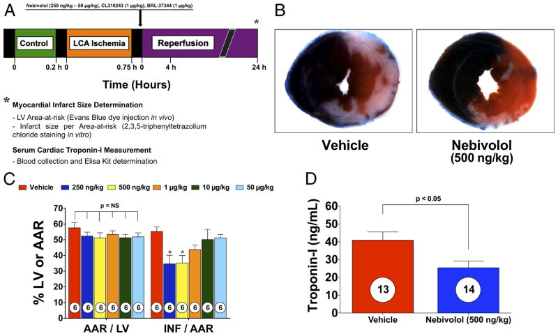
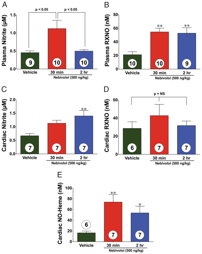
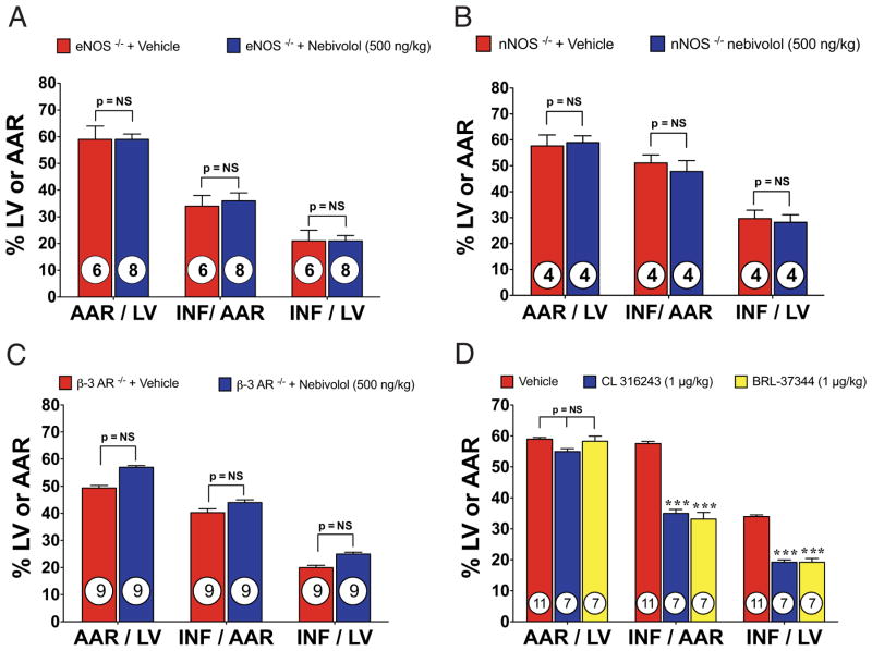
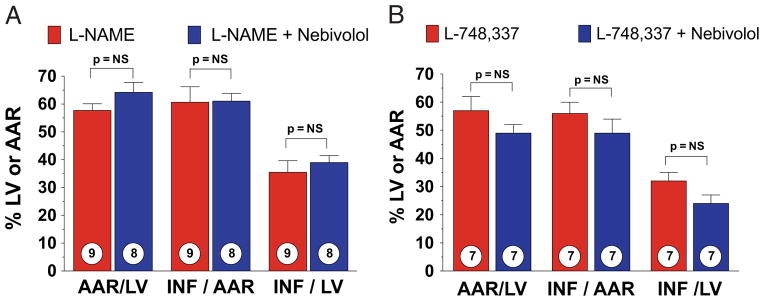
Methods: Mice were subjected to 45 min of myocardial ischemia in vivo followed by 24 h of reperfusion (R). Nebivolol (500 ng/kg), CL 316243 (1 μg/kg), BRL-37344 (1 μg/kg), or vehicle (VEH) was administered at the time of R. Myocardial area-at-risk (AAR) and infarct size (INF)/AAR was measured at 24 h of R. Cardiac tissue and plasma were collected to evaluate eNOS phosphorylation, neuronal nitric oxide synthase (nNOS), inducible nitric oxide synthase expression, and nitrite and nitrosothiol levels.

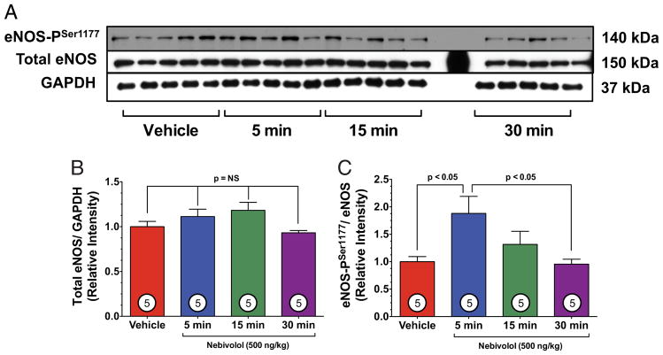
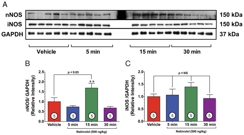
Results: Nebivolol (500 ng/kg) reduced INF/AAR by 37% (p < 0.001 vs. VEH) and serum troponin-I levels from 41 ± 4 ng/ml to 25 ± 4 ng/ml (p < 0.05 vs. VEH). CL 316243 and BRL-37344 reduced INF by 39% and 42%, respectively (p < 0.001 vs. VEH). Nebivolol and CL 316243 increased eNOS phosphorylation at Ser-1177 (p < 0.05 vs. VEH) and increased nitrite and total nitrosylated protein levels. Nebivolol and CL 316243 significantly increased myocardial nNOS expression. Nebivolol failed to reduce INF after MI/R in beta(3)-AR (-/-), eNOS(-/-), and in nNOS(-/-) mice.

Conclusions: Our results indicate that beta(3)-AR agonists protect against MI/R injury. Furthermore, the cardioprotective effects of beta(3)-AR agonists are mediated by rapid eNOS and nNOS activation and increased NO bioavailability.


Figures

Figure 1
Figure 1. Nebivolol Reduces Myocardial INF and Troponin-I
(A) Experimental protocol for this study. Mice were subjected to 45 min of left coronary artery (LCA) occlusion followed by 24 h of reperfusion. Myocardial area-at-risk (AAR) and myocardial infarct size (INF) were determined at 24 h of reperfusion. In addition, blood was collected at 24 h of reperfusion, and troponin-I was measured. Nebivolol or saline were injected directly into the left ventricular (LV) cavity just before reperfusion. (B) Representative mid-ventricular photomicrographs of mouse hearts after 45 min of myocardial ischemia and 24 h of reperfusion treated with vehicle (VEH) or nebivolol at the time of reperfusion. (C) Bar graph of myocardial AAR/LV and INF/AAR in mice treated with nebivolol. (D) Troponin levels in VEH and nebivolol-treated mice at 24 h after reperfusion. Nebivolol (500 ng/kg) administered at the time of reperfusion decreased serum troponin I levels, compared with VEH. Values are mean ± SEM. Numbers inside bars indicate the number of animals investigated in each group. *p < 0.05 versus VEH.
Figure 2
Figure 2. Nebivolol Increases eNOS-PSer1177
(A) Representative immunoblots of phosphorylation of endothelial nitric oxide synthase at serine residue 1177 (eNOS-PSer1177) and total endothelial nitric oxide synthase (eNOS) from the left ventricle of sham-operated mice receiving either vehicle (VEH) or nebivolol (500 ng/kg) via intracardiac injection at various time points. (B) Relative intensity of total eNOS remained constant compared with VEH. (C) Nebivolol increased the eNOS-PSer1177 at 15 min, compared with VEH (p < 0.05) and compared with 30 min (p < 0.05). Numbers inside bars indicate the number of animals studied in each group. GAPDH = glyceraldehyde-3-phosphate dehydrogenase.
Figure 3
Figure 3. Nebivolol Increases nNOS Expression
(A) Representative immunoblots of neuronal nitric oxide synthase (nNOS) and inducible nitric oxide synthase (iNOS) from the left ventricle of sham-operated mice receiving either VEH or nebivolol (500 ng/kg) via intracardiac injection at various time points. (B) Relative intensity of total nNOS increases after 15 min, compared with VEH (p < 0.05). **p < 0.01 versus 5 min and 30 min time points. (C) Relative intensity of total iNOS remained constant compared with VEH. Numbers inside bars indicate the number of animals studied in each group. Abbreviations as in Figure 2.
Figure 4
Figure 4. Nebivolol Increases NO Metabolites
Plasma and cardiac nitric oxide (NO) metabolites (nitrite and plasma total nitrosylated protein [RXNO]) measured after intracardiac injection of either vehicle (VEH) or nebivolol in sham-operated control mice. (A) Nitrite levels (μmol/l) increased in the plasma of nebivolol-treated mice. (B) The RXNO levels (nmol/l) increased after 30 min of nebivolol administration and remained elevated after 2 h. (C) Nitrite levels (μmol/l) increased in the heart of nebivolol-treated mice. (D) The RXNO levels in the heart did not significantly increase after 30 min of nebivolol administration. (E) The NO-Heme levels (nmol/l) significantly increased after 30 min of nebivolol administration and slightly decreased after 2 h. Values are mean ± SEM. *p < 0.05; **p < 0.01; numbers inside bars indicate the number of animals/group.
Figure 5
Figure 5. Nebivolol-Mediated Cardioprotection Is Lost in Pathway-Specific KO Mice
(A) Myocardial INF in eNOS−/− (eNOS knockout [KO]) mice treated with nebivolol (500 ng/kg). (B) Myocardial INF in nNOS−/− mice treated with nebivolol (500 ng/kg) at reperfusion. Bar graph of myocardial AAR/LV and INF/AAR in mice treated with nebivolol. (C) Myocardial INF in beta3-AR−/− mice treated with nebivolol. Treatment with nebivolol (500 ng/kg) at the time of reperfusion failed to reduce INF/AAR or INF/LV. (D) Myocardial INF in mice treated with CL 316243 (1 μg/kg) or BRL-37344 (1 μg/kg). No significant differences were observed with respect to the AAR/LV. Both CL 316243 and BRL 37344 significantly reduced myocardial INF/AAR and/LV. Values are mean ± SEM. ***p < 0.001; numbers inside bars indicate the number of animals/group. Abbreviations as in Figures 1, 2, and 3.
Figure 6
Figure 6. Nebivolol-Mediated Cardioprotection Is Lost With Pathway-Specific Pharmacological Inhibition
(A) Myocardial INF in wild-type mice treated with nitro-L-arginine methyl ester (L-NAME) (25 mg/kg) and mice treated with L-NAME (25 mg/kg) and nebivolol (500 ng/kg) at reperfusion. (B) Myocardial INF in wild-type mice treated with L-748,337 (100 μg/kg) and mice treated with L-748,337 (100 μg/kg) and nebivolol (500 ng/kg). Values are mean ± SEM. Numbers inside bars indicate the number of animals/group. Abbreviations as in Figure 1.

Comment in

References

    1. Bolli R. Mechanism of myocardial “stunning. Circulation. 1990;82:723–38. - PubMed
    1. Ovize M, Baxter GF, Di Lisa F, et al. Postconditioning and protection from reperfusion injury: where do we stand? Position paper from the Working Group of Cellular Biology of the Heart of the European Society of Cardiology. Cardiovasc Res. 2010;87:406–23. - PubMed
    1. Heusch G. Postconditioning: old wine in a new bottle? J Am Coll Cardiol. 2004;44:1111–2. - PubMed
    1. Hjalmarson A, Herlitz J, Holmberg S, et al. The Goteborg Metoprolol Trial. Effects on mortality and morbidity in acute myocardial infarction. Circulation. 1983;67:I26–32. - PubMed
    1. Lohse MJ, Engelhardt S, Eschenhagen T. What is the role of beta-adrenergic signaling in heart failure? Circ Res. 2003;93:896–906. - PubMed

Publication types

MeSH terms