. 2012 Jan 20;422(2):402-12.
doi: 10.1016/j.virol.2011.11.003. Epub 2011 Dec 5.

Characterization of a monoclonal anti-capsid antibody that cross-reacts with three major primate lentivirus lineages

Affiliations

Characterization of a monoclonal anti-capsid antibody that cross-reacts with three major primate lentivirus lineages

Brigitte E Sanders-Beer et al. Virology. .

Abstract

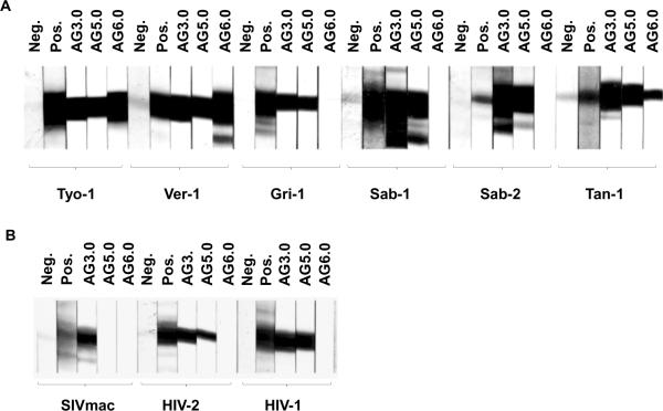
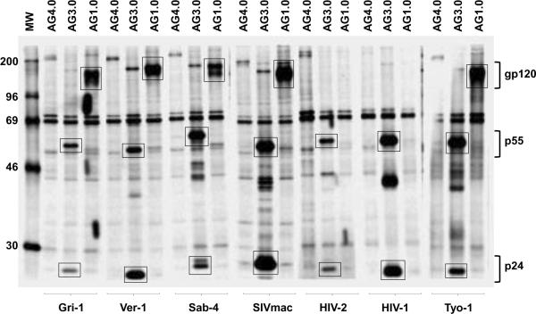
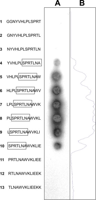
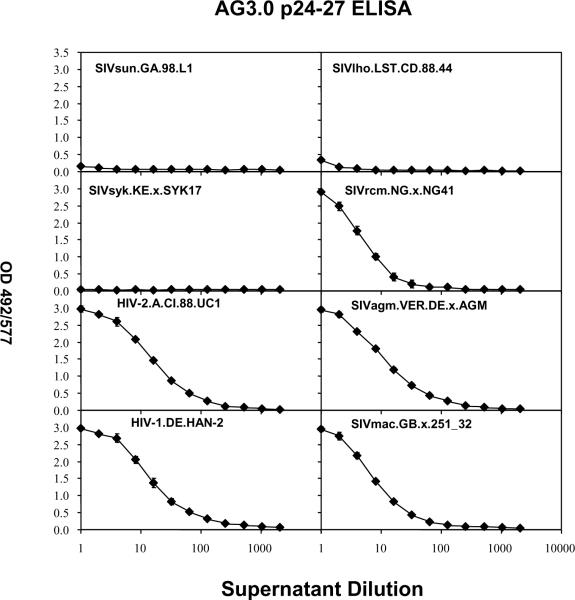
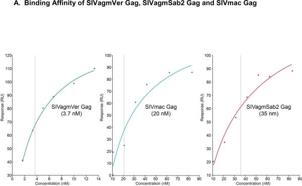
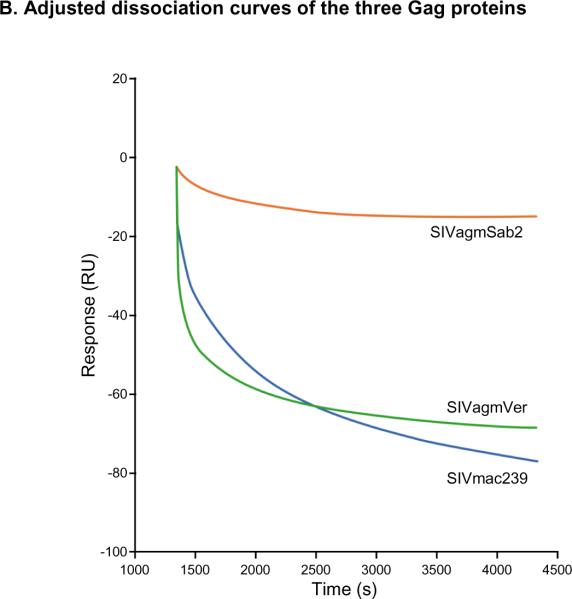
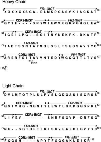
Mouse monoclonal antibodies with varying specificities against the Gag capsid of simian and human immunodeficiency virus (SIV/HIV) were generated by immunizing mice with whole inactivated SIVagmTYO-1. Monoclonal antibody AG3.0 showed the broadest reactivity recognizing the Gag capsid protein (p24-27) and Gag precursors p38, p55, and p150 of HIV-1, HIV-2, SIVmac, and SIVagm. Using overlapping peptides, the AG3.0 epitope was mapped in capsid to a sequence (SPRTLNA) conserved among HIV-1, HIV-2, SIVrcm, SIVsm/mac, and SIVagm related viruses. Because of its broad cross-reactivity, AG3.0 was used to develop an antigen capture assay with a lower detection limit of 100 pg/ml HIV-1 Gag p24. Interestingly, AG3.0 was found to have a faster binding on/off rate for SIVagmVer and SIVmac Gag than for SIVagmSab Gag, possibly due to differences outside the SPRTLNA motif. In addition, the ribonucleic acid (RNA) coding for AG3.0 was sequenced to facilitate the development of humanized monoclonal antibodies.


Figures

Figure 1
Figure 1. Western blot analysis
Reactivity of different Gag-specific monoclonal antibodies against various purified isolates of SIV and HIV in western blot performed as described in the materials and methods. A: Isolates from the four subspecies of African green monkeys. B: Isolates from rhesus macaques and humans. Neg: Negative control sera from uninfected individuals of the relevant species. Pos: Positive polyclonal control sera from individuals of the relevant species infected with the corresponding virus.
Figure 2
Figure 2. Radioimmunoprecipitation analysis
Binding of the AG3.0 monoclonal antibody to radiolabeled Gag protein from cells infected with various SIV and HIV isolates. The monoclonal antibodies AG1.0, recognizing gp120 from SIVagm and SIVmac, and AG4.0, recognizing a cell protein, were included as controls. Bands showing reactivity with p24–27, p55, and gp120 are marked with a box.
Figure 3
Figure 3. Fine epitope mapping of SIVmac Gag
Binding of the AG3.0 monoclonal antibody to synthetic peptides spanning the putative epitope in Gag within the sequence GGNYVHLPLSPRTLNAWVKLIEEKK (SIVmac251 Gag aa 141–165). The peptides, 13-mers with a 12 amino acid overlap were synthesized directly onto a cellulose membrane and binding of AG3.0 visualized by enhanced chemiluminescence (A). A density plot along the membrane was carried out to aid analysis or reactivity (B).
Figure 4
Figure 4. Antigen capture ELISA using various isolates
Supernatants from cells infected with various isolates of SIV and HIV were tested at different dilutions for reactivity in the AG3.0 based antigen capture assay as described in the material and methods.
Figure 5
Figure 5. Surface plasmon resonance (SPR) analysis
A: Binding affinity profiles of Gag proteins derived from different SIVs to AG3.0 measured by BIAcore as described in the materials and methods. Sensor chips coated in AG3.0 were exposed to purified preparations of the different Gag proteins at various concentrations. Binding was measured as a change in RU at equilibrium. The apparent kD values were determined using the BiaEvaluation software (GE Healthcare). B: Dissociation curves for the different Gag proteins (1.5 μg/ml) using an injection period of 1,080 sec, a dissociation time of 3,000 sec, and a flow rate of 5 μl/min.
Figure 6
Figure 6. Amino acid sequences of the heavy and light chains of the AG3.0 monoclonal antibody
The four conserved framework regions (FR1–FR4) and the three hypervariable complementarity determining regions (CDR1–CDR3) are displayed according to the IMGT nomenclature (Lefranc, 2007). Unoccupied amino acid positions are marked with a dash (−). X = positions in the conserved FR1 region of the heavy chain, at which the identity of the amino acid could not be unequivocally determined from the sequence.

References

    1. Aghokeng AF, Ayouba A, Mpoudi-Ngole E, Loul S, Liegeois F, Delaporte E, Peeters M. Extensive survey on the prevalence and genetic diversity of SIVs in primate bushmeat provides insights into risks for potential new cross-species transmissions. Infect Genet Evol. 2010;10(3):386–96. - PMC - PubMed
    1. Aghokeng AF, Bailes E, Loul S, Courgnaud V, Mpoudi-Ngolle E, Sharp PM, Delaporte E, Peeters M. Full-length sequence analysis of SIVmus in wild populations of mustached monkeys (Cercopithecus cephus) from Cameroon provides evidence for two co-circulating SIVmus lineages. Virology. 2007;360(2):407–18. - PMC - PubMed
    1. Allan JS, Short M, Taylor ME, Su S, Hirsch VM, Johnson PR, Shaw GM, Hahn BH. Species-specific diversity among simian immunodeficiency viruses from African green monkeys. J Virol. 1991;65(6):2816–28. - PMC - PubMed
    1. Allan JS, Whitehead EM, Strout K, Short M, Kanda P, Hart TK, Bugelski PJ. Strong association of simian immunodeficiency virus (SIVagm) envelope glycoprotein heterodimers: possible role in receptor-mediated activation. AIDS Res Hum Retroviruses. 1992;8(12):2011–20. - PubMed
    1. Bailes E, Gao F, Bibollet-Ruche F, Courgnaud V, Peeters M, Marx PA, Hahn BH, Sharp PM. Hybrid origin of SIV in chimpanzees. Science. 2003;300(5626):1713. - PubMed

Publication types

Associated data