Review
. 2012 Jan;22(1):33-41.
doi: 10.1016/j.tcb.2011.10.004. Epub 2011 Dec 9.

Multiple degradation pathways regulate versatile CIP/KIP CDK inhibitors

Affiliations
Review

Multiple degradation pathways regulate versatile CIP/KIP CDK inhibitors

Natalia G Starostina et al. Trends Cell Biol. 2012 Jan.

Abstract

The mammalian CIP/KIP family of cyclin-dependent kinase (CDK) inhibitors (CKIs) comprises three proteins--p21(Cip1/WAF1), p27(Kip1), and p57(Kip2)--that bind and inhibit cyclin-CDK complexes, which are key regulators of the cell cycle. CIP/KIP CKIs have additional independent functions in regulating transcription, apoptosis and actin cytoskeletal dynamics. These divergent functions are performed in distinct cellular compartments and contribute to the seemingly contradictory observation that the CKIs can both suppress and promote cancer. Multiple ubiquitin ligases (E3s) direct the proteasome-mediated degradation of p21, p27 and p57. This review analyzes recent data highlighting our current understanding of how distinct E3 pathways regulate subpopulations of the CKIs to control their diverse functions.


Figures

Box 1 Figure
Box 1 Figure
Figure 1
Figure 1. Diverse cellular functions of the CIP/KIP CKIs
A diagram showing primary functions of p21, p27, and p57 in regulating the cell cycle, transcription factors, the actin cytoskeleton, and apoptosis. Boxes are color coded, with light green reflecting functions that occur in the nucleus and magenta reflecting functions that occur in the cytoplasm. formula image is an inhibitory symbol, and an arrow is an activating symbol. See text for details.
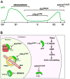
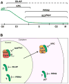
Figure 2
Figure 2. Ubiquitin ligase (E3) pathways that target the degradation of p21Cip1
A) A diagram of the p21 protein level (green line) through the cell cycle, starting from G0 phase, with brackets denoting the intervals of the cell cycle in which particular E3s regulate p21 levels. While many cells have low levels of p21 during G0 phase (shown), some cells express high levels of p21 in G0 phase (see references in [71]). B) A diagram showing the context of nuclear and cytoplasmic E3–p21 interactions. In the nucleus MDM2/MDMX targets the degradation of p21 in the context of both p21 and MDM2/MDMX binding to the 14-3-3τ protein. SCFSkp2 (with co-factor Cks1) targets p21 that is bound to cyclin–CDK2 and is phosphorylated on Ser130. CRL4Cdt2 targets p21 bound to chromatin-associated PCNA and phosphorylated on Ser114. In the cytoplasm, CRL2LRR1 targets p21 that inhibits ROCK to inactivate the RhoA–ROCK–LIMK pathway, which negatively regulates ADF/cofilin and actin cytoskeleton remodeling. APC/CCdc20 targets p21 for degradation at prometaphase via recognition of the p21 D-box motif. Note that APC/CCdc20 is active during mitosis when the nuclear envelope has disassembled and therefore nucleoplasm and cytoplasm are mixed. All E3s may not function within the same cells. See text for details
Figure 3
Figure 3. Ubiquitin ligase (E3) pathways that target the degradation of p27Kip1
A) A diagram of the p27 protein level (green line) through the cell cycle, initiating from G0 phase, with brackets denoting the intervals of the cell cycle in which particular E3s regulate p27 levels. The dashed line for E6-AP denotes the absence of information on when it is active during the cell cycle. B) A diagram showing the context of nuclear and cytoplasmic E3–p27 interactions. In the nucleus, SCFSkp2 targets p27 that is bound to CDK2–cyclin and is phosphorylated on Thr187. E6-AP targets nuclear p27, but the context is not known (e.g., is p27 bound to another protein or modified by a PTM?); denoted by a question mark. PIRH2 targets p27 in both the nucleus and the cytoplasm, but the context is not known. KPC targets p27 that has been translocated to the cytoplasm from the nucleus. One mechanism of translocation that is linked to KPC-mediated degradation is the binding of nuclear p27 to cyclin D2–CDK4/6, which is translocated to the cytoplasm in response to the phosphorylation of cyclin D2. All E3s may not function within the same cells. See text for details.

Similar articles

Cited by

References

    1. Murray AW. Recycling the cell cycle: cyclins revisited. Cell. 2004;116:221–234. - PubMed
    1. Besson A, et al. CDK inhibitors: cell cycle regulators and beyond. Dev Cell. 2008;14:159–169. - PubMed
    1. Glickman MH, Ciechanover A. The ubiquitin-proteasome proteolytic pathway: destruction for the sake of construction. Physiol Rev. 2002;82:373–428. - PubMed
    1. Sherr CJ, Roberts JM. CDK inhibitors: positive and negative regulators of G1-phase progression. Genes Dev. 1999;13:1501–1512. - PubMed
    1. Abbas T, Dutta A. p21 in cancer: intricate networks and multiple activities. Nat Rev Cancer. 2009;9:400–414. - PMC - PubMed

Publication types

Substances