Case Reports
. 2012 May;18(4):332-8.
doi: 10.1016/j.parkreldis.2011.11.019. Epub 2011 Dec 6.

First neuropathological description of a patient with Parkinson's disease and LRRK2 p.N1437H mutation

Affiliations
Case Reports

First neuropathological description of a patient with Parkinson's disease and LRRK2 p.N1437H mutation

Andreas Puschmann et al. Parkinsonism Relat Disord. 2012 May.

Abstract

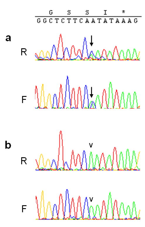
The c.4309A>C mutation in the LRRK2 gene (LRRK2 p.N1437H) has recently been reported as the seventh pathogenic LRRK2 mutation causing monogenic Parkinson's disease (PD). So far, only two families worldwide have been identified with this mutation. By screening DNA from seven brains of PD patients, we found one individual with seemingly sporadic PD and LRRK2 p.N1437H mutation. Clinically, the patient had levodopa-responsive PD with tremor, and developed severe motor fluctuations during a disease duration of 19 years. There was severe and painful ON-dystonia, and severe depression with suicidal thoughts during OFF. In the advanced stage, cognition was slow during motor OFF, but there was no noticeable cognitive decline. There were no signs of autonomic nervous system dysfunction. Bilateral deep brain stimulation of the subthalamic nucleus had unsatisfactory results on motor symptoms. The patient committed suicide. Neuropathological examination revealed marked cell loss and moderate alpha-synuclein positive Lewy body pathology in the brainstem. There was sparse Lewy pathology in the cortex. A striking finding was very pronounced ubiquitin-positive pathology in the brainstem, temporolimbic regions and neocortex. Ubiquitin positivity was most pronounced in the white matter, and was out of proportion to the comparatively weaker alpha-synuclein immunoreactivity. Immunostaining for tau was mildly positive, revealing non-specific changes, but staining for TDP-43 and FUS was entirely negative. The distribution and shape of ubiquitin-positive lesions in this patient differed from the few previously described patients with LRRK2 mutations and ubiquitin pathology, and the ubiquitinated protein substrate remains undefined.


Conflict of interest statement

Conflict of Interest:

The authors declare that they have no conflict of interest.

Figures

Figure 1
Figure 1. Sequencing results of LRRK2 exon 30
Heterozygosity for c.4309A>C (a, arrow) indicating a p.N1437H substitution, is seen in the forward (F) and reverse (R) sequencing reaction in DNA from the patient described here, but not in another patient (b, arrowhead).
Figure 2
Figure 2. Brain MRI imaging
T2-weighted imaging at 68 years of age revealed marked subcortical hyperintensities bilaterally. These spared the U-fibres and were unusually widespread, taking into consideration that the patient did not have diabetes, hypertension, or any other risk factor for small vessel disease. Top row, transverse sections, bottom row, coronal sections. The subthalamic nucleus (arrow in inset) was hyperintense and distinguishable with unusual contrast from the surrounding white matter. Slice thickness was 1 or 2 mm for the slices shown.
Figure 3
Figure 3. Macroscopic pathology
The formalin-fixed brain weighed 1420 grams and the aspect of the whole brain was without atrophy of cerebrum or cerebellum. The basal blood vessels showed only minimal signs of atheromatosis. After sectioning, there was an impression of mild diffuse thinning of the cortical ribbon. The substantia nigra was severely depigmented bilaterally, more severely on the left side. Both DBS electrode tracts could be followed to the subthalamic nucleus. The right DBS electrode tract went through the lateral thalamic nucleus, and in the lateral thalamic segment, there was an infarct of 1.5×1mm. Macroscopic pathology revealed only slight thinning of the cortical ribbon on coronar section. No widening of sulci was noted over the convexities. Substantia nigra was depigmented (not shown). The cortical entrances of the DBS electrodes are visible in the left upper picture.
Figure 4
Figure 4. Brainstem Neuropathology
a and b) Substantia nigra (SN), H&E: There was almost total loss of melanin-containing neurons of substantia nigra pars compacta (SNpc) bilaterally. Only a few clusters of pigment-containing neurons were seen in the most ventromedial parts of as well as towards its latero-dorsal end. The few remaining SN cells showed atrophy and loss of pigmentation. A few Lewy bodies were seen (LB). c) Locus coeruelus, H&E: Marked cell loss and LB pathology. d) SN, alpha-synuclein: LBs and isolated dot-like structures stained positively for alpha-synuclein. e) SN, ubiquitin: Pronounced ubiquitin pathology with dense spheroids and dot-like structures as well as irregularly shaped foamy inclusions. f) White matter (crus cerebri adjacent to SN), ubiquitin: multitudinous ubiquitin-positive structures in the form of small dots, larger speroids and intermediate cloud-like positivities. Magnification: a) x125; b–c) x400; d–f) x200; insert in e) x400, enlarged.
Figure 5
Figure 5. Cortical Neuropathology
a–d) Parietal cortex, e–g) Entorhinal cortex. a and e) Alpha-synuclein: very sparse cortical alpha-synuclein pathology with single Lewy bodies (a) or Lewy neurites (e) per field. b and f) Tau: sparse tau-positive lesions of unspecific shape. c,d and g) Ubiquitin Marked ubiquitin positive pathology in the parietal cortex and very numerous lesions in the entorhinal cortex, including dense spheroids, less dense granular lesions with irregular shapes. h) White matter subjacent to entorhinal cortex, ubiquitin: Very high burden of ubiquitin-positive lesions. Magnification: a–c) and e–h) x200; d) and inserts in g) x400.

References

    1. Ross OA, Soto-Ortolaza AI, Aasly JO, Abahuni N, Annesi G, Bacon JA, et al. LRRK2 exonic variants and susceptibility to Parkinson s disease. Lancet Neurol. 2011 Oct;10(10):898–908. - PMC - PubMed
    1. Zimprich A, Biskup S, Leitner P, Lichtner P, Farrer M, Lincoln S, et al. Mutations in LRRK2 cause autosomal-dominant parkinsonism with pleomorphic pathology. Neuron. 2004 Nov 18;44(4):601–7. - PubMed
    1. Wider C, Dickson DW, Wszolek ZK. Leucine-rich repeat kinase 2 gene-associated disease: redefining genotype-phenotype correlation. Neurodegener Dis. 2010;7(1–3):175–9. - PMC - PubMed
    1. Paisan-Ruiz C, Jain S, Evans EW, Gilks WP, Simon J, van der Brug M, et al. Cloning of the gene containing mutations that cause PARK8-linked Parkinson's disease. Neuron. 2004 Nov 18;44(4):595–600. - PubMed
    1. Aasly JO, Vilarino-Guell C, Dächsel JC, Webber PJ, West AB, Haugarvoll K, et al. Novel pathogenic LRRK2 p.Asn1437His substitution in familial Parkinson's disease. Mov Disord. 2010 Oct 15;25(13):2156–63. - PMC - PubMed

Publication types