Review
. 2012 Feb 1;52(3):556-592.
doi: 10.1016/j.freeradbiomed.2011.11.002. Epub 2011 Nov 12.

Role of reactive oxygen and nitrogen species in the vascular responses to inflammation

Affiliations
Review

Role of reactive oxygen and nitrogen species in the vascular responses to inflammation

Peter R Kvietys et al. Free Radic Biol Med. .

Abstract

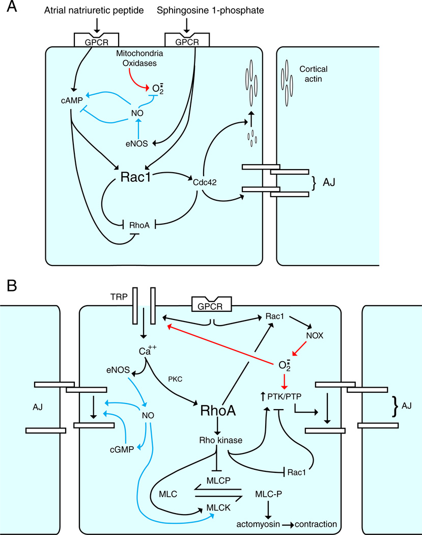
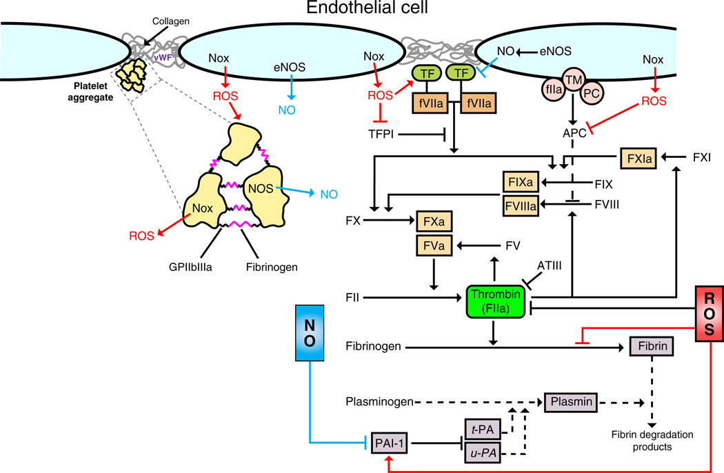
Inflammation is a complex and potentially life-threatening condition that involves the participation of a variety of chemical mediators, signaling pathways, and cell types. The microcirculation, which is critical for the initiation and perpetuation of an inflammatory response, exhibits several characteristic functional and structural changes in response to inflammation. These include vasomotor dysfunction (impaired vessel dilation and constriction), the adhesion and transendothelial migration of leukocytes, endothelial barrier dysfunction (increased vascular permeability), blood vessel proliferation (angiogenesis), and enhanced thrombus formation. These diverse responses of the microvasculature largely reflect the endothelial cell dysfunction that accompanies inflammation and the central role of these cells in modulating processes as varied as blood flow regulation, angiogenesis, and thrombogenesis. The importance of endothelial cells in inflammation-induced vascular dysfunction is also predicated on the ability of these cells to produce and respond to reactive oxygen and nitrogen species. Inflammation seems to upset the balance between nitric oxide and superoxide within (and surrounding) endothelial cells, which is necessary for normal vessel function. This review is focused on defining the molecular targets in the vessel wall that interact with reactive oxygen species and nitric oxide to produce the characteristic functional and structural changes that occur in response to inflammation. This analysis of the literature is consistent with the view that reactive oxygen and nitrogen species contribute significantly to the diverse vascular responses in inflammation and supports efforts that are directed at targeting these highly reactive species to maintain normal vascular health in pathological conditions that are associated with acute or chronic inflammation.


Figures

Fig. 1
Fig. 1
Representative endothelial-dependent relaxing factors (EDRFs) and endothelial-dependent hypopolarizing factors (EDHFs). Endothelial activation by shear stress or ligands of G-protein-coupled receptors (GPCRs) increases intracellular levels of Ca2+, which is the initial event in the generation of EDRFs and EDHFs. EDRFs (red pathways): elevated endothelial Ca2+ levels activate at least two enzymes that generate EDRFs, nitric oxide synthase (eNOS) and cyclo-oxygenase (COX). NO (or HNO) derived from eNOS diffuses to smooth muscle and activates cGMP. The prostanoid PGI2 derived from COX interacts with its receptor (IPR) and activates cAMP. These second-messenger systems, in turn, activate Ca2+-dependent K+ channels (BKCa++) in smooth muscle resulting in inhibition of voltage-gated Ca2+ channels (Ca++V). The resultant decrease in intracellular Ca2+ leads to smooth muscle relaxation. EDHFs (green pathways): elevated endothelial Ca2+ levels also result in smooth muscle hyperpolarization by either activating Ca2+-dependent K+ channels or generating H2O2. Increased K+ efflux (via SKCa++ or IKCa++) results in endothelial membrane hyperpolarization (MHP), which can be transmitted to smooth muscle via myoendothelial gap junctions. Alternatively, the K+ ions entering the internal elastic lamina can cause smooth muscle hyperpolarization. In either case, Ca++V are inhibited and smooth muscle Ca2+ levels decrease, resulting in smooth muscle relaxation. Finally, various oxidases (e.g., NADPH oxidase, COX) as well as eNOS (e.g., uncoupled) can generate superoxide, which is rapidly converted to H2O2 by Cu,Zn-SOD. H2O2 can then diffuse to the smooth muscle where it activates BKCa++ and inhibits Ca++V.
Fig. 2
Fig. 2
Leukocyte recruitment to sites of injury/infection: dominant role of ROS. (A) Activation of macrophages by infection/injury. PAMPs (e.g., LPS) generated by infection and DAMPs (e.g., HMGB1) generated by stressed/necrotic cells serve as ligands for TLR and RAGE on interstitial sentinel cells (e.g., macrophages). Ligation of these receptors results in activation of NADPH oxidase (NOX), which generates ROS (superoxide and its dismutation product H2O2) (red pathway). ROS can be (1) exported into the interstitium to affect adjacent endothelial cells and (2) further activate NOX (e.g., via an NF-κB pathway) leading to the generation of additional ROS (feed-forward mechanism). Activation of NF-κB can also increase iNOS levels, which results in NO generation (blue pathway). NO can dampen the inflammatory response by interacting with superoxide within macrophages or adjacent cells. (B) Rapid and delayed phases of endothelial activation. Endothelial cells are activated by the proinflammatory milieu (chemokines, cytokines, ROS, LPS, HMGB1). Rapid activation (NF-κB independent) of endothelial cells by chemokines and ROS results in further ROS generation via NOX (red pathways). Endothelial ROS contribute to adhesion molecule expression (P-selectin), which facilitates leukocyte rolling. ROS have also been implicated in endothelial cell generation of leukocyte activators (e.g., PAF and CXCL8), which are sequestered within the glycocalyx and facilitate leukocyte adhesion to the endothelium. Leukocyte adhesion to endothelium results in the clustering of endothelial adhesion molecules (docking structures). The resultant cell signaling disrupts adherens junctions (AJ) via NOX-derived ROS and facilitates leukocyte TEM. Delayed activation (NF-κB dependent) of endothelium reinforces the leukocyte–endothelial adhesive interactions via continued and amplified generation of ROS, chemokines, and cytokines via the NF-κB pathway.
Fig. 3
Fig. 3
Role of Rac1/RhoA balance in endothelial barrier integrity: impact of ROS and NO. (A) Basal permeability. In quiescent endothelial cells, cytosolic Rac1 is dominant and inhibits RhoA activation. Rac1/Cdc42 stabilizes both the cortical actin and the adherens junction components (VE-cadherin/β-catenin). Basal eNOS activity generates NO (blue pathway), which dampens any superoxide production within endothelial cells. In addition, low levels of NO increase cAMP, whereas high levels decrease cAMP. Agents that tend to strengthen barrier integrity can either increase cAMP, leading to an increase in the Rac1/RhoA ratio (e.g., ANP), or increase eNOS activity (e.g., S1P). (B) Increased permeability. In response to proinflammatory mediators (e.g., VEGF, thrombin, histamine), RhoA becomes dominant and inhibits cytosolic Rac1. Activation of GPCRs increases intracellular Ca2+ levels (via TRP channels) and activates RhoA. RhoA (via Rho kinase) (1) increases the PTK/PTP ratio, leading to disorganization of adherens junctions, and (2) activates MLCK and inhibits MLCP, leading to actomyosin-mediated contraction. Paradoxically Rho kinase activates Rac1 at the membrane, leading to activation of NADPH oxidase and superoxide production (red pathway), which in turn increases the PTK/PTP ratio. The increased intracellular Ca2+ also activates eNOS. Although low levels of NO (not shown) tend to stabilize adherens junctions, higher levels of NO (shown) favor disruption of the junctions as well as increasing MLCK activity (blue pathways).
Fig. 4
Fig. 4
Cooperative role of ROS and NO in angiogenesis. (A) Generation of VEGF by macrophages. Both ROS (red pathways) and NO (blue pathways) generated during hypoxia/inflammation can decrease PHD activity, which activates the HIF pathway as well as the NF-κB pathway. The HIF pathway leads to transcription of VEGF. The NF-κB pathway leads to transcription of inflammatory cytokines. During the inflammatory response the NF-κB predominates, and during the resolution of the inflammatory response the HIF pathway predominates. NO modulates both pathways at several points, being stimulatory in the HIF pathway and inhibitory in the NF-κB pathway. (B) Initiation of angiogenesis: endothelial destabilization. VEGF derived from macrophages or recruited endothelial progenitor cells (EPC) interacts with VEGF receptor (VEGFR) on selected endothelial “tip” cells to initiate cell migration. VEGFR ligation activates both eNOS and NOX to generate NO and superoxide, respectively. NO (blue pathways) is involved in (1) loosening of the adherens junctions and (2) MMP activation and TIMP inhibition to facilitate degradation of the ECM. Superoxide (red pathways) is involved in (1) formation of lamellipodia and (2) cyclic alterations in the strength of focal adhesion complexes (FACs). The migratory tip cells are followed by hyperpermeable (destabilized junctions) stalk cells.
Fig. 5
Fig. 5
Role of ROS and NO in coagulation and platelet aggregation. Thrombus formation involves the adhesion, activation, and aggregation of platelets as well as activation of the coagulation cascade. With vessel injury, platelets bind to exposed collagen and von Willebrand factor (vWF). Upon activation, platelets bind to one another using fibrinogen and GPIIb/IIIa to form an aggregate. Tissue factor (TF) triggers coagulation by binding to activated factor VII (fVII), which ultimately leads to activation of other coagulation factors and the conversion of prothrombin to thrombin. Thrombin cleaves fibrinogen to generate fibrin monomers, which polymerize to form a stable clot. Fibrinolysis (proteolytic degradation of fibrin), which prevents excess thrombus growth, is mediated by plasminogen and its activators (t-PA and u-PA) and controlled by plasminogen activator inhibitor-1 (PAI-1), which inhibits t-PA and u-PA. ROS and NO are known to modulate the coagulation pathway and fibrinolysis by interacting with multiple components of this cascade. ROS (red pathways) can promote the initiation of coagulation by targeting the TF–fVII complex as well as tissue factor protein inhibitor (TFPI). ROS also promote coagulation and thrombus formation by inhibiting the production of activated protein C (APC), enhancing the conversion of fibrinogen to thrombin and enhancing PAI-1 activity. NO (blue pathways) tends to exert an opposite effect on the coagulation cascade. In addition, NO targets the platelets to inhibit aggregation, whereas ROS promote this process. NADPH oxidase (Nox) seems to be a major endothelial cell source of the ROS that modulate platelet aggregation.

References

    1. Granger DN, Senchenkova E. Inflammation and the microcirculation. In: Granger DN, Granger JP, editors. Colloquium Series in Integrated Systems Physiology: from Molecules to Function. Princeton, NJ: Morgan-Claypool Life. Sci; 2010. - PubMed
    1. Ley K. The microcirculation in inflammation. In: Tuma RDW, Ley K, editors. Handbook of Physiology: Microcirculation. San Diego: Academic Press; 2008. pp. 387–448.
    1. Majno G. Role of inflammatory mediators in angiogenesis. Am. J. Pathol. 1998;153:1035–1039. - PMC - PubMed
    1. Ryan GB, Majno G. Acute inflammation: a review. Am. J. Pathol. 1977;86:183–276. - PMC - PubMed
    1. Esmon C. Crosstalk between inflammation and thrombosis. Maturitas. 2004;47:305–314. - PubMed

Publication types

Substances

LinkOut - more resources