. 2012 Mar 15;180(2-3):230-6.
doi: 10.1016/j.resp.2011.11.012. Epub 2011 Dec 2.

Persistence of the nasotrigeminal reflex after pontomedullary transection

Affiliations

Persistence of the nasotrigeminal reflex after pontomedullary transection

W Michael Panneton et al. Respir Physiol Neurobiol. .

Abstract

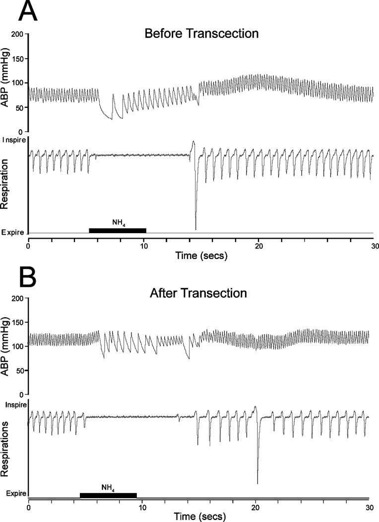
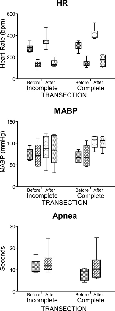
Most behaviors have numerous components based on reflexes, but the neural circuits driving most reflexes rarely are documented. The nasotrigeminal reflex induced by stimulating the nasal mucosa causes an apnea, a bradycardia, and variable changes in mean arterial blood pressure (MABP). In this study we tested the nasotrigeminal reflex after transecting the brainstem at the pontomedullary junction. The nasal mucosae of anesthetized rats were stimulated with ammonia vapors and their brainstems then were transected. Complete transections alone induced an increase in resting heart rate (HR; p<0.001) and MABP (p<0.001), but no significant change in ventilation. However, the responses to nasal stimulation after transection were similar to those seen prior to transection. HR still dropped significantly (p<0.001), duration of apnea remained the same, as did changes in MABP. Results from rats whose transection were incomplete are discussed. These data implicate that the neuronal circuitry driving the nasotrigeminal reflex, and indirectly the diving response, is intrinsic to the medulla and spinal cord.


Figures

Figure 1
Figure 1
Photomicrographs of the rat brainstem illustrating the pontomedullary transections and some presumptive pathways destroyed. The brainstem was cut near the pontomedullary junction (A, shaded area) through the trapezoid body but spared the ventral third of the spinal trigeminal tract (A, unshaded boxed area), allowing primary afferent fibers innervating paranasal areas to proceed caudally. Only brains with a complete transection of their ventral surface were considered transected (B); note the spared ventrolateral corners with their spinal trigeminal tracts beneath (B, arrows). A dorsal view of a transected brainstem (C) shows the absence of major blood clots, since the tool used spared major blood vessels. Horizontal sections through a transected brainstem shows complete transection ventrally between the caudal superior olivary nucleus and the rostral the facial nucleus most ventrally (D), approximately 800µm dorsal to horizontal zero (E), and 1.5mm dorsal to horizontal zero (F). Arrow in D shows the spared trapezoid body, the arrow in E shows the spared spinal trigeminal tract, while that in F shows the complete transection. Such transections destroyed descending fibers originating in the dorsolateral pons (an injection of biotinylated dextran amine into cardiorespiratory areas of the peribrachial complex is shown in G). Labeled fibers form this injection descended medial to the exiting fibers of the seventh nerve (H); these fibers were transected. Immunostained sections with antibodies to tyrosine hydroxylase suggest that rostral neurons of the catecholamine A5 cell group were also destroyed (I), but more caudally placed A5 neurons probably survived. Abbreviations: A5, A5 catecholamine neuron group; KF, Kölliker-Füse nucleus; LVe, lateral vestibular nucleus; Mo5, motor trigeminal nucleus; PB, parabrachial complex; PnC, pontine reticular nucleus, pars caudalis; Pr5, principal sensory trigeminal nucleus; SO, superior olivary nucleus; SubC, subcoeruleus nucleus; Tz, nucleus of the trapezoid body; VCA, ventral cochlear nucleus, pars anterior; VLL, ventral nucleus of the lateral lemniscus; bc, brachium conjunctivum; icp, inferior cerebellar peduncle; py, pyramidal tract; sp5, spinal trigeminal tract; tz, trapezoid body; 7n, facial nerve; 8n, vestibular nerve.
Figure 2
Figure 2
Tracings illustrating the cardiorespiratory responses to nasal stimulation with ammonia vapors (black bar) for 5 sec both before (A) and after (B) pontomedullary transection. Note that normal respirations were not altered by such transections, that the apnea and bradycardia induced were similar before and after transection, but resting arterial blood pressure (ABP) was elevated by the cut.
Figure 3
Figure 3
Box plots illustrating the changes induced in heart rate, mean arterial blood pressure, and apnea after stimulating the nasal mucosa of rats with ammonia vapors both before and after transecting the brainstem at the pontomedullary junction. Transections judged Incomplete (n=7) are shown on the left while those judged Complete (n=6) are on the right. Mean bradycardia induced by nasal stimulation was similar despite degree of transection, but resting heart rate increased after transection, especially complete transections. Mean arterial blood pressure stayed relatively the same after ammonia stimulation, but resting pressures were increased after complete transections. The mean time for apnea induced by nasal stimulation was unchanged with transections, but became more variable. Lighter shades of boxes represent control data prior to nasal stimulation while darker shades represent cardiorespiratory consequences during nasal stimulation with ammonia vapors. See text for levels of significance.

References

    1. Alheid GF, Milsom WK, McCrimmon DR. Pontine influences on breathing: an overview. Resp. Physiol. Neurobiol. 2004;143:105–114. - PubMed
    1. Angell James JE, de Burgh Daly M. Reflex respiratory and cardiovascular effects of stimulation of receptors in the nose of the dog. J. Physiol. (Lond.) 1972;220:673–696. - PMC - PubMed
    1. Blix AS, Folkow B. Cardiovascular adjustments to diving in mammals and birds. In: Sheperd JT, Abboud FM, editors. Handbook of Physiology - The Cardiovascular System. Bethesda, MD: American Physiological Society; 1983. pp. 917–945.
    1. Butler PJ, Jones DR. The comparative physiology of diving in vertebrates. Adv. Comp. Physiol. Biochem. 1982;8:179–364. - PubMed
    1. Butler PJ, Jones DR. Physiology of diving of birds and mammals. Physiol. Rev. 1997;77:837–899. - PubMed

Publication types

MeSH terms