

The expression and function of vascular endothelial growth factor in retinal pigment epithelial (RPE) cells is regulated by 4-hydroxynonenal (HNE) and glutathione S-transferaseA4-4
- PMID: 22155253
- PMCID: PMC3259230
- DOI: 10.1016/j.bbrc.2011.11.113
The expression and function of vascular endothelial growth factor in retinal pigment epithelial (RPE) cells is regulated by 4-hydroxynonenal (HNE) and glutathione S-transferaseA4-4
Abstract
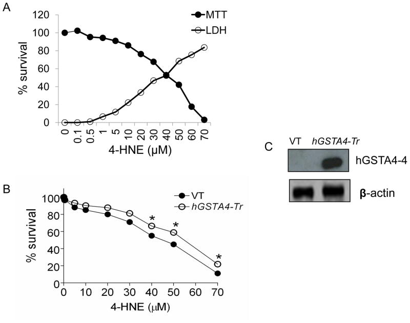
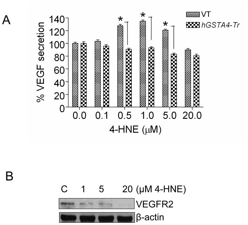
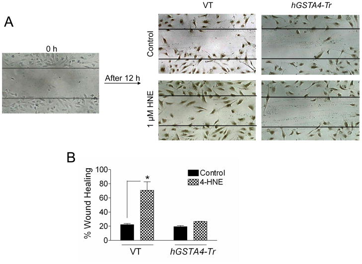
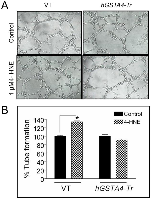
It is well established that 4-hydroxynonenal (HNE) plays a major role in oxidative stress-induced signaling and the toxicity of oxidants. Surprisingly our recent studies also demonstrate that low levels of HNE generated during oxidative stress promote cell survival mechanisms and proliferation. Since the expression and secretion of VEGF is known to be affected by Oxidative stress, during present studies, we have examined dose dependent effect of HNE on VEGF expression and secretion in a model of retinal pigment epithelial (RPE) cells in culture. Results of these studies showed that while inclusion of 0.1 μM HNE in the medium caused increased secretion of VEGF, its secretion and expression was significantly suppressed in the presence of >5 μM HNE in the media. These concentration dependent hormetic effects of HNE on VEGF secretion could be blocked by the over expression of GSTA4-4 indicating that these effects were specifically attributed to HNE and regulated by GSTA4-4. VEGF secreted into the media showed angiogenic properties as indicated by increased migration and tube formation of HUVEC in matrigel when grown in media from RPE cells treated with 1 μM HNE. The corresponding media from GSTA4-4 over expressing RPE cells had no effect on migration and tube formation of HUVEC in matrigel. These results are consistent with earlier studies showing that at low concentrations, HNE promotes proliferative mechanisms and suggest that HNE induces VEGF secretion from RPE cells that acts in a paracrine fashion to induce angiogenic signaling mechanism in the endothelial cells. These findings may suggest a role of HNE and GSTA4-4 in oxidative stress induced proliferative retinopathies.
Copyright © 2011 Elsevier Inc. All rights reserved.
Figures
References
-
- Chung FL, Chen HJ, Nath RG. Lipid peroxidation as a potential endogenous source for the formation of exocyclic DNA adducts. Carcinogenesis. 1996;17:2105–2111. - PubMed
-
- Yancopoulos GD, Davis S, Gale NW, et al. Vascular-specific growth factors and blood vessel formation. Nature. 2000;407:242–248. - PubMed
-
- Adamis AP, Shima DT, Yeo KT, et al. Synthesis and secretion of vascular permeability factor/vascular endothelial growth factor by human retinal pigment epithelial cells. Biochem Biophys Res Commun. 1993;193:631–638. - PubMed
Publication types
MeSH terms
Substances
Grants and funding
LinkOut - more resources
Full Text Sources
