

Differential ascending projections of temporomandibular joint-responsive brainstem neurons to periaqueductal gray and posterior thalamus of male and female rats
- PMID: 22155654
- PMCID: PMC3273606
- DOI: 10.1016/j.neuroscience.2011.11.042
Differential ascending projections of temporomandibular joint-responsive brainstem neurons to periaqueductal gray and posterior thalamus of male and female rats
Abstract
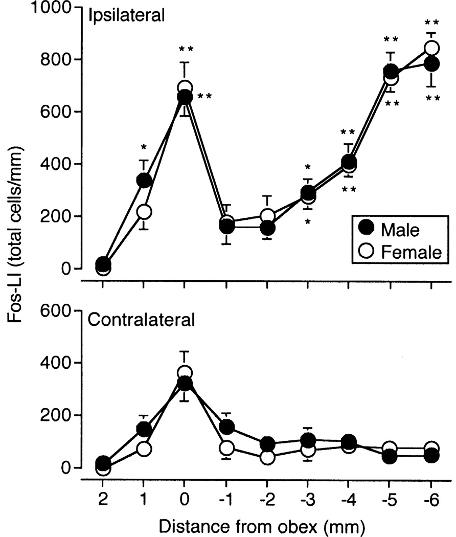
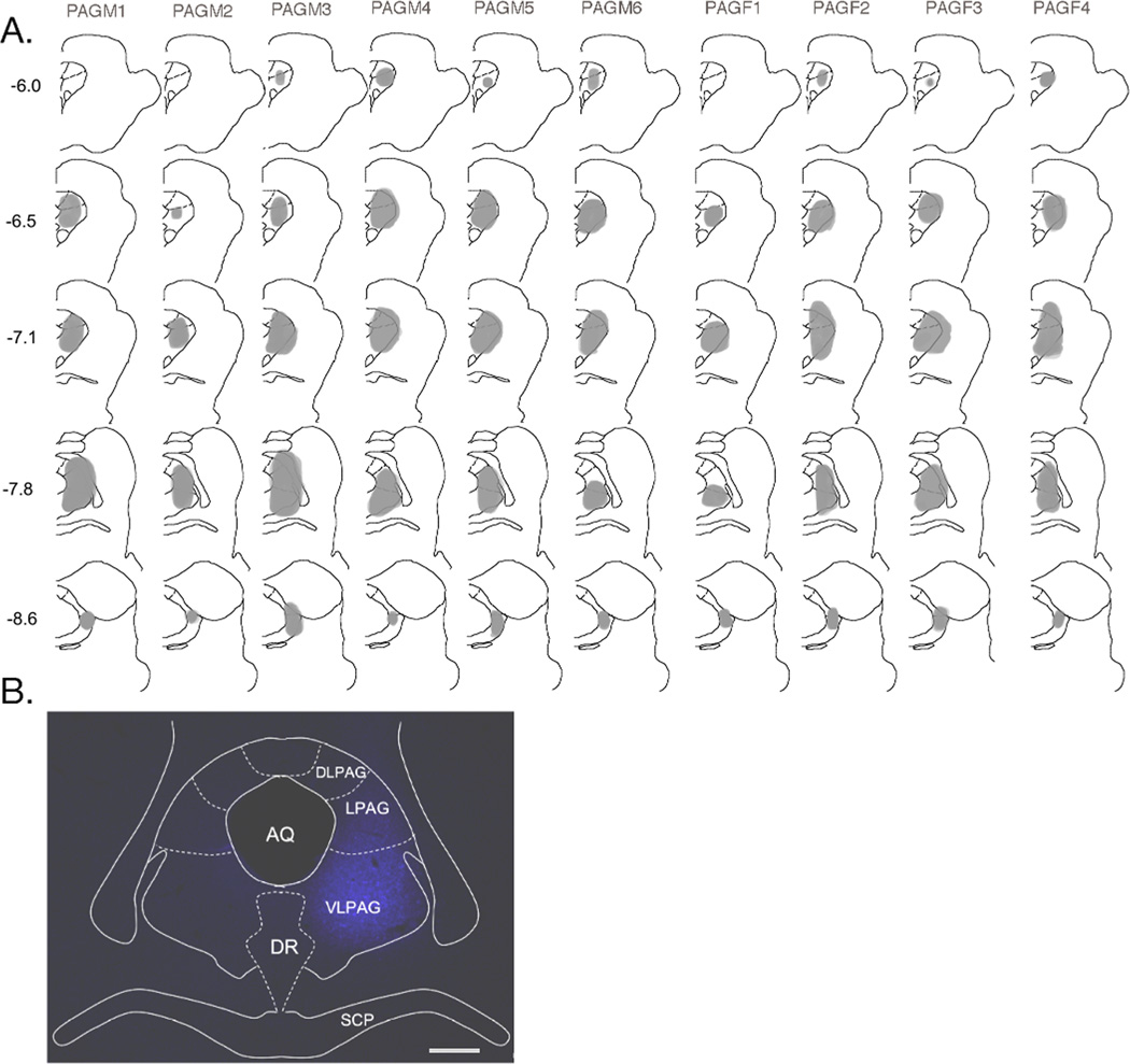
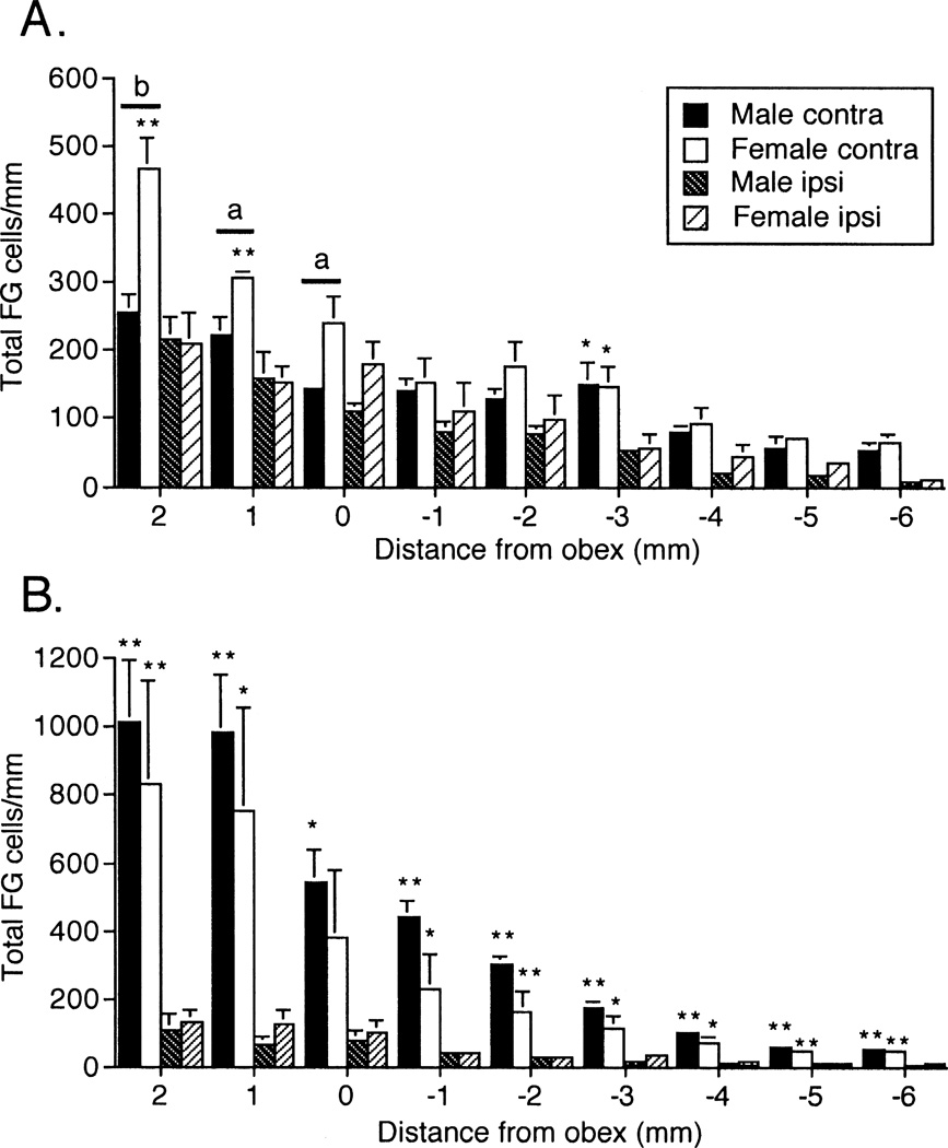
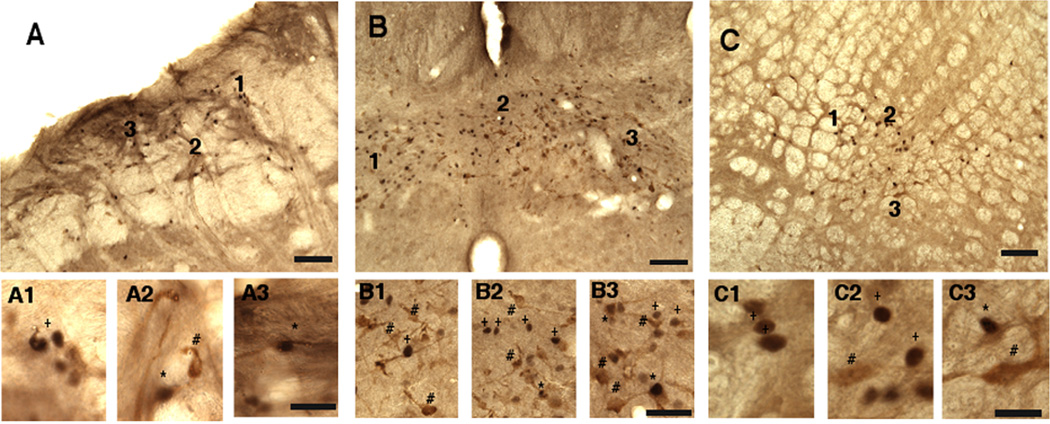
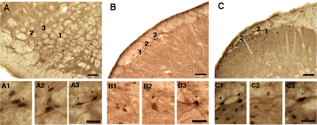
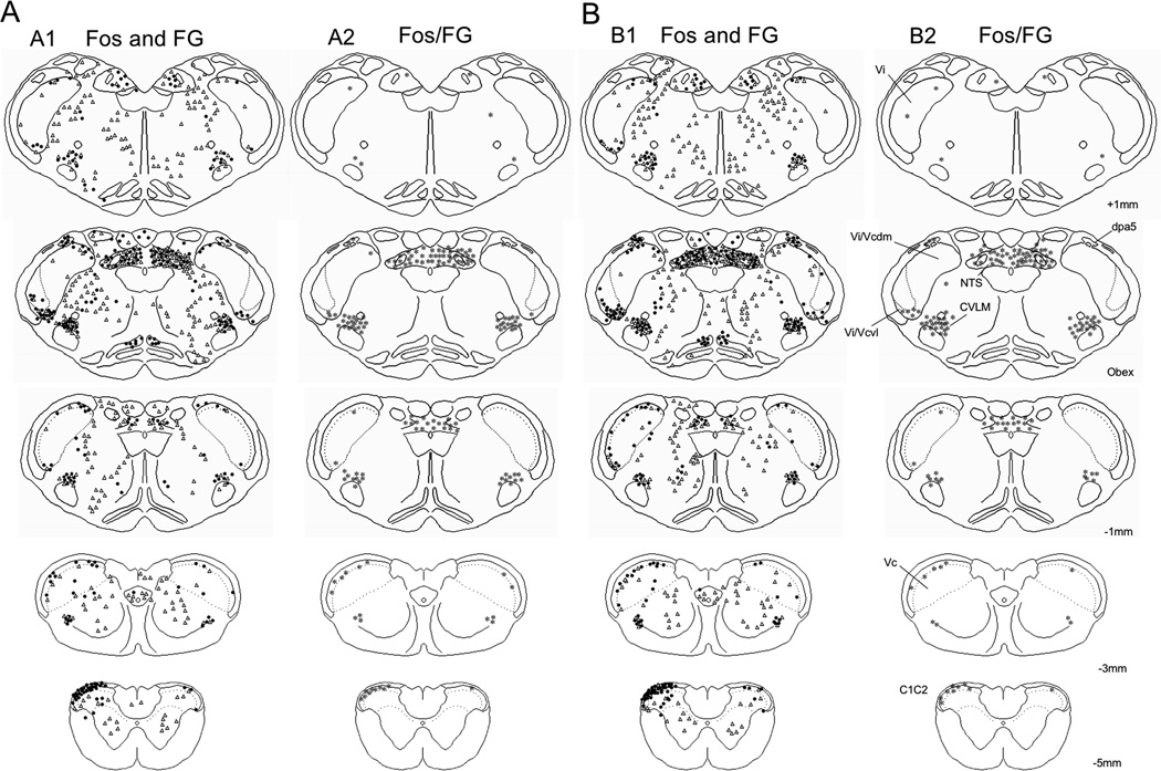
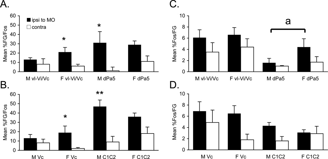
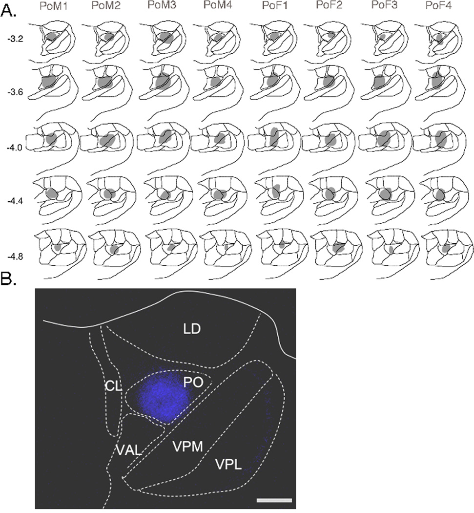
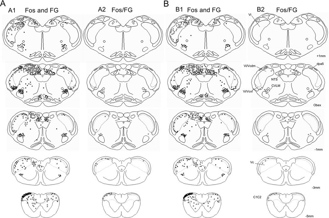
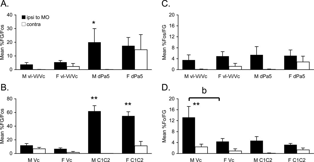
Several craniofacial pain conditions, including temporomandibular joint disorders (TMJDs), are more prevalent in women than men. The basis for sex differences in deep craniofacial pain is not known. The present study compared the magnitude of ascending projections from temporomandibular joint (TMJ)-responsive neurons in trigeminal brainstem with the ventrolateral periaqueductal gray (vlPAG) or posterior nucleus of the thalamus (Po) in males and female rats. Fluorogold (FG) was injected into vlPAG or Po, and TMJ-responsive neurons were identified by Fos-like immunoreactivity (Fos-LI) after mustard oil injection. TMJ-evoked Fos-LI was similar in males and females; however, significant differences in cell counts were seen for FG single-labeled and Fos/FG double-labeled neurons in trigeminal brainstem. After vlPAG injections, the number of FG-labeled neurons in trigeminal subnucleus interpolaris (Vi), ventral interpolaris/caudalis transition (vl-Vi/Vc), and dorsal paratrigeminal region (dPa5) was greater in females than males. The percentage of Fos/FG double-labeled neurons in vl-Vi/Vc and dPa5 after vlPAG injection also was greater in females than males. In contrast, after Po injections, males displayed a greater number of FG-labeled neurons in superficial laminae (Lam I/II) of trigeminal subnucleus caudalis (Vc) and upper cervical spinal cord (C(1-2)) and deeper laminae (Lam III/V) at C(1-2) than females. The percentage of Fos/FG double-labeled neurons in Lam I/II of Vc after Po injection also was greater in males than females. These data revealed significant sex differences in ascending projections from TMJ-responsive neurons in trigeminal brainstem. Such differences may influence the ability of males and females to recruit autonomic reflexes and endogenous pain control circuits relevant for TMJ nociception.
Copyright © 2011 IBRO. Published by Elsevier Ltd. All rights reserved.
Figures
References
-
- Ajika K, Krulich L, Fawcett CP, McCann SM. Effects of estrogen on plasma and pituitary gonadotropins and prolactin, and on hypothalamic releasing and inhibiting factors. Neuroendocrinology. 1972;9:304–315. - PubMed
-
- Bandler R, Shipley MT. Columnar organization in the midbrain periaqueductal gray: modules for emotional expression? Trends Neurosci. 1994;17:379–389. - PubMed
-
- Beitz AJ. The organization of afferent projections to the midbrain periaqueductal gray of the rat. Neuroscience. 1982;7:133–159. - PubMed
-
- Bereiter DA. Sex differences in brainstem neural activation after injury to the TMJ region. Cells Tissues Organs. 2001;169:226–237. - PubMed
-
- Bereiter DA, Bereiter DF. Morphine and NMDA receptor antagonism reduce c-fos expression in spinal trigeminal nucleus produced by acute injury to the TMJ region. Pain. 2000;85:65–77. - PubMed
Publication types
MeSH terms
Grants and funding
LinkOut - more resources
Full Text Sources
