. 2012 Feb 1;590(3):631-46.
doi: 10.1113/jphysiol.2011.224477. Epub 2011 Dec 12.

Systemic cholecystokinin amplifies vago-vagal reflex responses recorded in vagal motor neurones

Affiliations

Systemic cholecystokinin amplifies vago-vagal reflex responses recorded in vagal motor neurones

Edouard Viard et al. J Physiol. .

Abstract

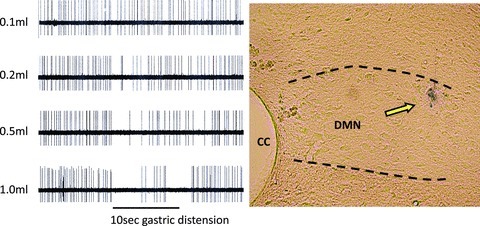
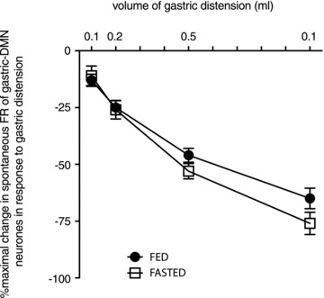
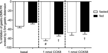
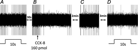
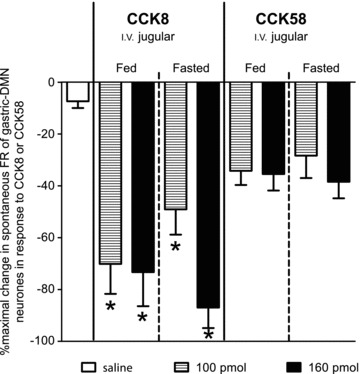
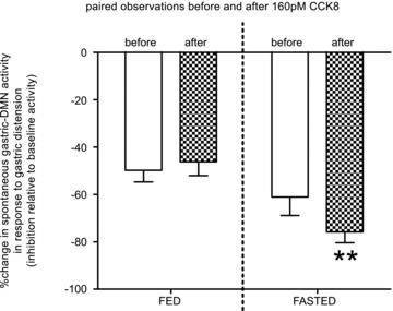
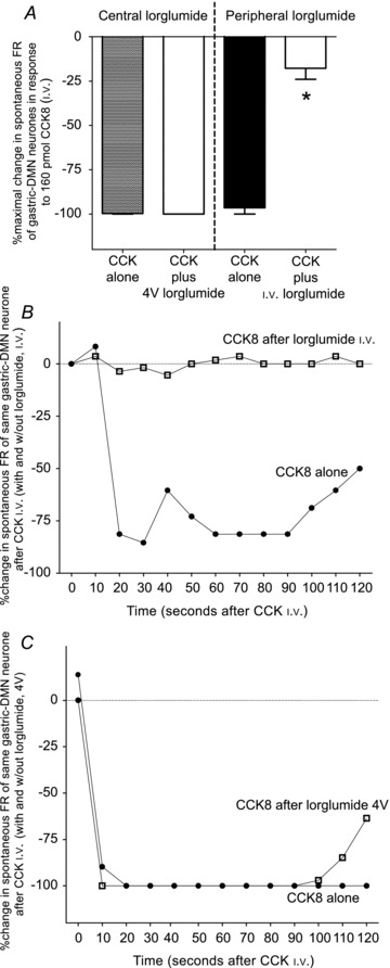
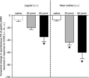
Cholecystokinin (CCK) is a potent regulator of visceral functions as a consequence of its actions on vago-vagal reflex circuit elements. This paper addresses three current controversies regarding the role of CCK to control gastric function via vago-vagal reflexes. Specifically: (a) whether CNS vs. peripheral (vagal afferent) receptors are dominant, (b) whether the long (58) vs. short (8) isoform is more potent and (c) whether nutritional status impacts the gain or even the direction of vago-vagal reflexes. Our in vivo recordings of physiologically identified gastric vagal motor neurones (gastric-DMN) involved in the gastric accommodation reflex (GAR) show unequivocally that: (a) receptors in the coeliac-portal circulation are more sensitive in amplifying gastric vagal reflexes; (b) in the periphery, CCK8 is more potent than CCK58; and (c) the nutritional status has a marginal effect on gastric reflex control. While the GAR reflex is more sensitive in the fasted rat, CCK amplifies this sensitivity. Thus, our results are in stark contrast to recent reports which have suggested that vago-vagal reflexes are inverted by the metabolic status of the animal and that this inversion could be mediated by CCK within the CNS.


Figures

Figure 1
Figure 1
Left panel: raw oscillograph records of the inhibition of spontaneous activity of DMN neurone responding to different volumes of gastric (antral) distension. Over 95% of physiologically identified DMN neurones are inhibited by antral distension (i.e. gastro-excitatory DMN neurones that are inhibited by distension; McCann & Rogers, 1992)). Antral distension volumes of 0.1, 0.2, 0.5 and 1.0 ml in the emptied stomachs of either previously ‘fed’ or ‘fasted’ rats caused a distension-locked reduction in spontaneous activity of the gastric-DMN neurones involved in the gastric accommodation reflex in a distension-volume-dependent manner. Right panel: histological verification of microelectrode recording site. CC, central canal; DMN, dorsal motor nucleus of vagus.
Figure 2
Figure 2. Gastric distension inhibits spontaneous DMN activity in a volume-dependent fashion
Spontaneous FR activity of DMN neurones is relatively constant. Gastric distension via balloon expansion (0.1–1.0 ml volumes) caused incremental increases in the inhibition of the spontaneous FR of DMN neurones. Basal FR for each individual DMN neurone was considered 100% and then compared to relative amount of FR inhibition caused by each level of distension. Thus, each neurone served as its own control. The resultant stimulus/response curves generated by fed vs. fasted preparations were analysed for best fit of non-linear regression curves with intercept through zero. Fasted animals demonstrated a slight increase in sensitivity to gastric distension at the higher volumes relative to animals that had not been fasted. Non-linear regression analysis of these curves indicate different slopes (‘fed’=−75.6 ± 3.4; ‘fasted’=−87.8 ± 4.1; F1,242= 5.329; P= 0.02).
Figure 3
Figure 3. Effect of systemic CCK8 or CCK58 (1nmol, i.v.) on the sensitivity of DMN neurones to gastric distension
As also seen in Figs 1 and 2, under basal conditions, the spontaneous FR of gastro-excitatory-DMN neurones was reduced approximately 50% in response to 0.5 ml antral distension, regardless of the metabolic status of the animal. In this subset of experiments, fed or fasted animals received intravenous (i.v.) delivery of 1 nmol of either CCK8 or CCK58. Two-way analysis of variance revealed a main effect of the 1 nmol CCK treatment (F2,139= 14.48; P < 0.0001), but no main effect of the metabolic state nor was there an interaction between these two sources of variance. Bonferroni post tests between basal and each of the two different CCK subtypes indicate that CCK8 amplifies the inhibition of gastric-DMN neurones to 0.5 ml antral distension in either the fed or fasted state (#P < 0.0001); in contrast CCK58 significantly amplifies this inhibition only in the fed state (†P < 0.05) relative to the basal responses.
Figure 4
Figure 4. Raw oscillographic record of a gastric-DMN neurone firing rate (FR) in response to antral distension before and after CCK8 i.v.
A, basal spontaneous FR of DMN neurone is transiently reduced during the 10 s antral distension (0.5 ml volume). B, after basal FR recovered, CCK8 (160 pmol in 3.3 μl total volume) was injected i.v. and evoked a pronounced suppression of spontaneous DMN activity. C, within 2–3 min after CCK8 i.v., spontaneous FR returns to a steady level. D, after intravenous CCK8, suppression of spontaneous FR of same gastric-DMN neurone evoked by 10 s antral distension for 10 s is amplified relative to the response elicited prior to CCK8 exposure.
Figure 5
Figure 5. Percentage maximal change in spontaneous FR of identified gastric-DMN neurones occurring within 2 min following 100 or 160 pmol i.v. injections of CCK8 or 58 (or 3.3 μl i.v. saline) in either fed or fasted animals
Comparable numbers of identified gastric-DMN neurones were sensitive to either CCK8 (29 neurones sensitive/32 neurones identified) or CCK58 (26 neurones sensitive/28 neurones identified). Two-way analysis of variance (metabolic state versus doses of CCK) revealed that there was no interaction between metabolic state of the animal or CCK doses. There was a main effect of CCK on spontaneous DMN firing rate (F4,63= 19.61; P < 0.0001) that is exclusively attributable to CCK8. Note, there was no main effect of metabolic state (F4,63= 0.13; P= 0.72).
Figure 6
Figure 6. Responses of identified gastric-DMN neurones to modest antral distension (0.5 ml) before and after systemic administration of CCK8 (160 pmol) in either fed or fasted animals were made as paired observations
Repeated measures two-way analysis of variance revealed that the matching effect of these paired observations was extremely significant (F17,17= 9.82; P < 0.0001) and there was a significant interaction between metabolic state and the presence of CCK8 (F1,17= 12.42; P= 0.0026); the effect of CCK to amplify the inhibition of gastric-DMN FR in response to modest gastric distension was more evident in the fasted animals. Given that the interaction is statistically significant, the P values of the CCK main effect (F1,17= 4.54; P= 0.048) and the metabolic main effect (F1,17= 6.23; P= 0.023) are more difficult to interpret. However, it can be readily appreciated that in the fasted animal, exogenous CCK (given systemically at approximate physiological doses) amplifies the inhibition of gastric-DMN firing rate in response to antral distension. That is, at physiological doses, exogenous, systemic CCK does not modulate the ‘fasted’ gastric accommodation reflex to resemble the reflex elicited in the ‘fed’ state.
Figure 7
Figure 7. Peripheral versus central antagonism of the effects of systemic CCK8 on DMN spontaneous activity
A, bar graph represents averaged maximal effects of CCK8 to inhibit spontaneous DMN firing rate with and without lorglumide administered either centrally or peripherally. Application of lorglumide to the 4V floor has no effect on the attenuation of the spontaneous activity of the gastric-DMN neurones evoked by systemic CCK8 in fasted rats. In contrast, i.v.-administered lorglumide reduced the attenuating effect of CCK8 on the spontaneous activity of the gastric-DMN neurones by approximately 80% (F3,16= 112; P < 0.0001). B, plot of the spontaneous activity of an identified gastric-DMN neurone exposed to CCK8 with and without i.v.-administered lorglumide. Note that the CCK8 effect is completely blocked after i.v. lorglumide. C, similar plot of the spontaneous activity of an identified gastric-DMN neurone exposed to CCK8 with and without 4V-administered lorglumide. Spontaneous activity of this gastric-DMN neurone was not affected following 4V application of lorglumide.
Figure 8
Figure 8. Percentage maximal change in spontaneous FR of identified gastric-DMN neurones occurring within 2 min following 0, 30, or 60 pmol injections of CCK8 into either the systemic (jugular (i.v.)) or coeliac/mesenteric (near coeliac (i.a.)) circulation of fasted animals
Two-way analysis of variance revealed that the route of administration was very significant (F1,36= 9.23; P= 0.0044) as was the dose of CCK8 (F2,36= 19.04; P < 0.0001). There was no interaction between these sources of variance. *P < 0.05 Bonferroni multiple comparisons to saline.

Similar articles

Cited by

References

    1. Andresen MC, Yang MY. Non-NMDA receptors mediate sensory afferent synaptic transmission in medial nucleus tractus solitarius. Am J Physiol Heart Circ Physiol. 1990;259:H1307–H1311. - PubMed
    1. Baptista V, Zheng ZL, Coleman FH, Rogers RC, Travagli RA. Cholecystokinin octapeptide increases spontaneous glutamatergic synaptic transmission to neurons of the nucleus tractus solitarius centralis. J Neurophysiol. 2005;94:2763–2771. - PMC - PubMed
    1. Berthoud HR, Patterson LM. Anatomical relationship between vagal afferent fibers and CCK-immunoreactive entero-endocrine cells in the rat small intestinal mucosa. Acta Anat (Basel) 1996;156:123–131. - PubMed
    1. Buelke-Sam J, Holson JF, Bazare JJ, Young JF. Comparative stability of physiological parameters during sustained anesthesia in rats. Lab Anim Sci. 1978;28:157–162. - PubMed
    1. Burdyga G, de Lartigue G, Raybould HE, Morris R, Dimaline R, Varro A, Thompson DG, Dockray GJ. Cholecystokinin regulates expression of Y2 receptors in vagal afferent neurons serving the stomach. J Neurosci. 2008;28:11583–11592. - PMC - PubMed

Publication types