. 2012 Feb;40(4):e29.
doi: 10.1093/nar/gkr1146. Epub 2011 Dec 7.

Characterization of DNA methyltransferase specificities using single-molecule, real-time DNA sequencing

Affiliations

Characterization of DNA methyltransferase specificities using single-molecule, real-time DNA sequencing

Tyson A Clark et al. Nucleic Acids Res. 2012 Feb.

Abstract

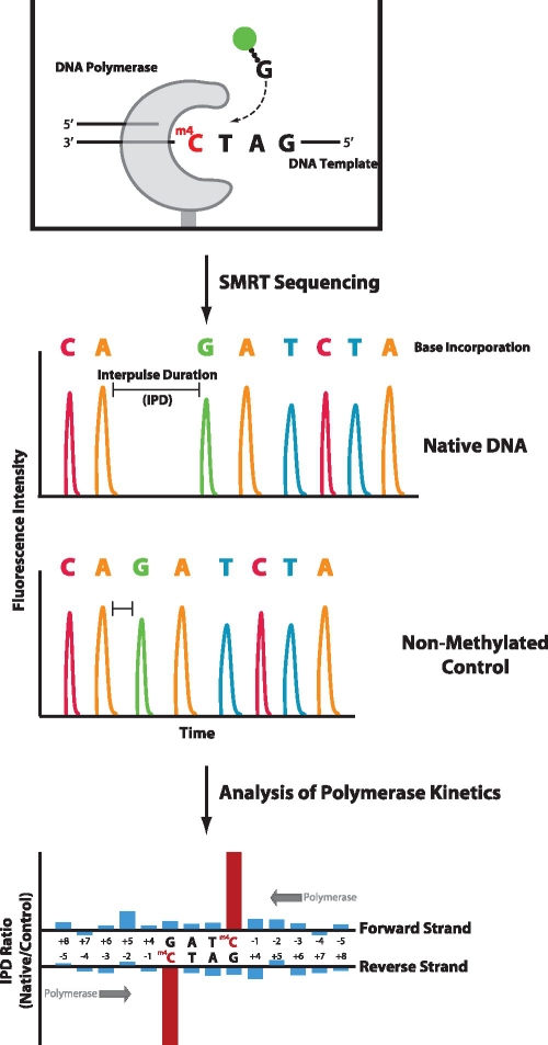
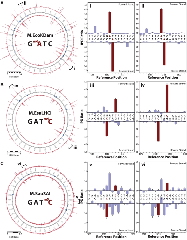
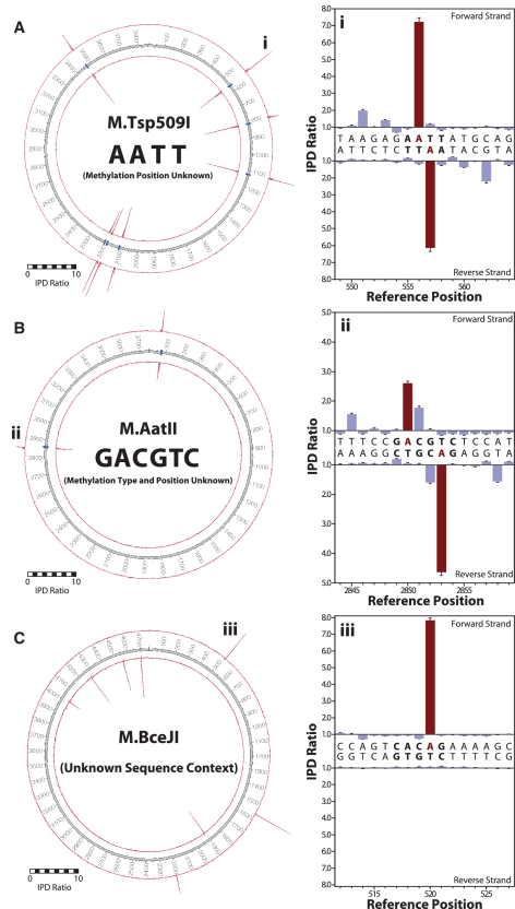
DNA methylation is the most common form of DNA modification in prokaryotic and eukaryotic genomes. We have applied the method of single-molecule, real-time (SMRT®) DNA sequencing that is capable of direct detection of modified bases at single-nucleotide resolution to characterize the specificity of several bacterial DNA methyltransferases (MTases). In addition to previously described SMRT sequencing of N6-methyladenine and 5-methylcytosine, we show that N4-methylcytosine also has a specific kinetic signature and is therefore identifiable using this approach. We demonstrate for all three prokaryotic methylation types that SMRT sequencing confirms the identity and position of the methylated base in cases where the MTase specificity was previously established by other methods. We then applied the method to determine the sequence context and methylated base identity for three MTases with unknown specificities. In addition, we also find evidence of unanticipated MTase promiscuity with some enzymes apparently also modifying sequences that are related, but not identical, to the cognate site.


Figures

Figure 1.
Figure 1.
Principle of characterizing MTase specificities by SMRT DNA sequencing. In SMRT sequencing, single molecules of an engineered phi29-based polymerase are monitored in real-time, using fluorescently-labeled phospholinked nucleotides, as they synthesize a complementary strand from the DNA template strand that contains methylated bases. The timing of fluorescence pulses corresponding to nucleotide incorporations is analyzed and compared with a control template lacking methylated bases. The kinetics of DNA synthesis is affected by the presence of a methylated base in the template, e.g. by increasing the time prior to nucleotide binding across the methylated base, resulting in an increased IPD. The ratio of IPDs between native and control samples for each template position yield kinetic signatures for identifying the presence of methylated bases in the DNA template, thus defining MTase specificities.
Figure 2.
Figure 2.
Confirmation of activity of cloned methyltransferase genes using methylation protection assays. Plasmids containing genes encoding the MTases (identified in the text above the gel images) were isolated from E. coli ER2796, a strain which lacks endogenous methyltransferase activities. Methylated plasmid DNAs (right lane in each case) were linearized using PstI then challenged with restriction endonucleases (REases) that are expected to be blocked by the action of the relevant cloned MTase. CviQI, RsaI, Sau3AI, Tsp509I, AatII, AluI, BstNI, NspI, HpaII and SacI samples were assayed using the equivalent cognate REases. EcoKDam and EsaLHCI samples were assayed using REase MboI, EsaBC1I using REase AluI, EcoKDcm with REase PspGI and EsaBC2I with REase SalI. Control PCR product DNAs (left lane in each case) were restricted with the same enzymes as the methylated plasmids but without PstI. The SacI unmodified (PCR product) DNA was also cleaved with PstI as this construct contains an additional PstI site within the coding sequence of the SacI MTase gene. M = 1 kb DNA-Ladder Marker.
Figure 3.
Figure 3.
MTase specificities determined from SMRT sequencing. The sequence context 5′-GTAC-3′ is methylated by (A) M.CviQI (m6A) and (B) M.RsaI (m4C). The left panel shows IPD ratio data for both strands over the entire plasmid, with the inner and outer circles representing the reverse and forward DNA template strands, respectively. Template positions are indicated by the numbered track middle circle, with blue markers denoting occurrences of MTase target sequence contexts. The right panels show IPD ratios for two representative template positions containing the target context (bold letters); the methylated base is highlighted in red. Error bars represent the standard error of the ratio of means and is calculated as described in the ‘Materials and Methods’ section.
Figure 4.
Figure 4.
MTase specificities determined from SMRT sequencing. The sequence context 5′-GATC-3′ is methylated by (A) M.EcoKDam (m6A), (B) M.EsaLHCI (m4C) and (C) M.Sau3AI (m5C). The left panel shows IPD ratio data for both strands over the entire plasmid, with the inner and outer circles representing the reverse and forward DNA template strands, respectively. Template positions are indicated by the numbered track middle circle, with blue markers denoting occurrences of MTase target sequence contexts. The right panels show IPD ratios for two representative template positions containing the target context (bold letters), the methylated base is highlighted in red. Error bars represent the standard error of the ratio of means and is calculated as described in the ‘Materials and Methods’ section.
Figure 5.
Figure 5.
Determination of unknown MTase specificities. Base-resolved MTase target specificities were resolved for (A) M.Tsp509I as 5′-A m6A TT-3′, (B) M.AatII as 5′-G m6A CGTC-3′ and (C) M.BceJI as 5′-CAC m6A G-3′. The left panel shows IPD ratio data for both strands over the entire plasmid, with the inner and outer circles representing the reverse and forward DNA template strands, respectively. Template positions are indicated by the numbered track middle circle, with blue markers denoting occurrences of MTase target sequence contexts (if known). The right panels show IPD ratios for two representative template positions containing the target context (bold letters), the methylated base is highlighted in red. Error bars represent the standard error of the ratio of means and is calculated as described in the ‘Materials and Methods’ section.
Figure 6.
Figure 6.
Sequence context specificity of M.EcoKDam. Kinetic signals for the target 5′-GATC-3′ and all 1-base neighboring sequence contexts containing adenine at the second position are compared. The values plotted represent average IPD ratios of all occurrences of each sequence context (error bars are s.e.m.). The inset shows an example for a methylation signal at the off-target 5′-GACC-3′ sequence context. Error bars represent the standard error of the ratio of means and is calculated as described in the ‘Materials and Methods’ section.

References

    1. Malone T, Blumenthal RM, Cheng X. Structure-guided analysis reveals nine sequence motifs conserved among DNA amino-methyltransferases, and suggests a catalytic mechanism for these enzymes. J. Mol. Biol. 1995;253:618–632. - PubMed
    1. Kumar S, Cheng X, Klimasauskas S, Mi S, Posfai J, Roberts RJ, Wilson GG. The DNA (cytosine-5) methyltransferases. Nucleic Acids Res. 1994;22:1–10. - PMC - PubMed
    1. Wu JC, Santi DV. Kinetic and catalytic mechanism of HhaI methyltransferase. J. Biol. Chem. 1987;262:4778–4786. - PubMed
    1. Roberts RJ, Vincze T, Posfai J, Macelis D. REBASE–a database for DNA restriction and modification: enzymes, genes and genomes. Nucleic Acids Res. 2010;38:D234–D236. - PMC - PubMed
    1. Marinus MG, Casadesus J. Roles of DNA adenine methylation in host-pathogen interactions: mismatch repair, transcriptional regulation, and more. FEMS Microbiol. Rev. 2009;33:488–503. - PMC - PubMed

Publication types

MeSH terms

Substances

Associated data