Review
. 2011 Dec 1;25(23):2436-52.
doi: 10.1101/gad.179184.111.

Mechanisms and functions of Tet protein-mediated 5-methylcytosine oxidation

Affiliations
Review

Mechanisms and functions of Tet protein-mediated 5-methylcytosine oxidation

Hao Wu et al. Genes Dev. .

Abstract

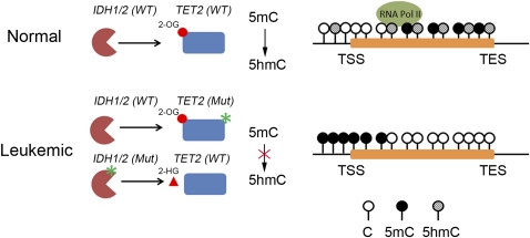
Ten-eleven translocation 1-3 (Tet1-3) proteins have recently been discovered in mammalian cells to be members of a family of DNA hydroxylases that possess enzymatic activity toward the methyl mark on the 5-position of cytosine (5-methylcytosine [5mC]), a well-characterized epigenetic modification that has essential roles in regulating gene expression and maintaining cellular identity. Tet proteins can convert 5mC into 5-hydroxymethylcytosine (5hmC), 5-formylcytosine (5fC), and 5-carboxylcytosine (5caC) through three consecutive oxidation reactions. These modified bases may represent new epigenetic states in genomic DNA or intermediates in the process of DNA demethylation. Emerging biochemical, genetic, and functional evidence suggests that Tet proteins are crucial for diverse biological processes, including zygotic epigenetic reprogramming, pluripotent stem cell differentiation, hematopoiesis, and development of leukemia. Insights into how Tet proteins contribute to dynamic changes in DNA methylation and gene expression will greatly enhance our understanding of epigenetic regulation of normal development and human diseases.


Figures

Figure 1.
Figure 1.
Domain architecture of mouse Tet proteins. Schematic diagrams of predicted functional domains in the mouse Tet proteins (Tet1–3). Three conserved domains—including CXXC zinc finger, the cysteine-rich region (Cys-rich), and the double-stranded β-helix (DSBH) fold of the 2OG-Fe(II) dioxygenase domain—are indicated. Numbers represent the amino acid numbers. Note that Tet2 does not contain a predicted CXXC domain. Multiple sequence alignment of the catalytic motif of the JBP/Tet family of dioxygenases. Sequences used in the alignment include the Trypanosoma brucei JBP1 (Q9U6M3) and JBP2 (Q57X81); human TET1 (Q8NFU7), TET2 (Q6N021), and TET3 (O43151); and mouse Tet1 (GU079948), Tet2 (GU079949), and Tet3 (Q8BG87). Numbers represent the amino acid numbers. Alignment was performed using MultAlin (http://bioinfo.genotoul.fr/multalin). The color code is defined by the MultAlin program. Predicted Fe(II)- and 2OG-binding sites are highlighted.
Figure 2.
Figure 2.
Detection methods of 5mC oxidation derivatives. (A) Schematic diagrams of in vitro biochemical assays followed by 2D TLC. The spots corresponding to 5mC and its oxidation products are indicated in a representative 2D TLC plate. (B) Flow chart of the procedures used for quantifying 5mC oxidation derivatives in mouse genomic DNA. Representative data of high-performance LC (HPLC) and LC-MS/MS assays are shown. (C) Schematic representations of how antibodies specific to 5mC oxidation products can be used in immunoprecipitation or immunostaining assays. (D) Schematic representation of the 5hmC glucosylation reaction catalyzed by β-glucosyltransferase (β-GT) of T-even bacteriophages.
Figure 3.
Figure 3.
Proposed models of Tet-initiated DNA demethylation pathways. DNA methylation (5mC) is established and maintained by DNA methyltransferases (Dnmt). In mammals, 5mC can be hydroxylated by the Tet family of dioxygenases to generate 5hmC. 5hmC is recognized poorly by Dnmt1 and can lead to replication-dependent passive demethylation. 5hmC can be further oxidized by Tet proteins to produce 5fC and 5caC. Alternatively, 5hmC may be further deaminated to become 5hmU by AID/APOBECC deaminases. 5hmU, 5fC, and 5caC can be excised from DNA by glycosylases such as TDG. A putative decarboxylase may directly convert 5caC to C. Cytosine and its derivatives are highlighted in red. DNA glycosylase TDG-catalyzed reactions are indicated by blue arrows.
Figure 4.
Figure 4.
Relationship of Tet1/5hmC and transcription in mouse ES cells. Schematic representation of the relative enrichment of Tet1 and major histone modifications (H3K4me3 and H3K27me3), as well as the distribution of 5mC/5hmC at four major classes of genes: (1) Highly transcribed genes with a CpG-rich promoter are associated with high levels of Tet1 and H3K4me3 at their promoters, as well as high levels of 5mC/5hmC within their gene bodies (particularly at 3′ intragenic regions). (2) Actively transcribed genes with CpG-poor promoters (e.g., a subset of pluripotency factors such as Nanog and Tcl1) are associated with medium/low levels of Tet1 and 5hmC at their promoters as well as within their gene bodies. (3) Polycomb-repressed yet “poised” genes (e.g., lineage-specific transcription factors) are enriched with high levels of Tet1, 5hmC, H3K4me3, and H3K27me3 at their extended promoters. (4) Silent genes with a CpG-poor promoter (e.g., tissue-specific genes) are generally associated with sparsely distributed 5mC and 5hmC at their proximal promoter, but are devoid of high levels of Tet1 and PRC2. (TSS) Transcriptional start site; (TES) transcriptional end site.
Figure 5.
Figure 5.
Dynamic changes of 5mC and 5hmC levels in the paternal and maternal genomes during preimplantation development. Tet3 is highly expressed in the oocyte and one-cell zygote. Immediately after fertilization, Tet3 may potentially relocate from the cytoplasm to the paternal nucleus to convert 5mC to 5hmC. Subsequently, paternal and maternal genomes undergo replication-dependent dilution of 5hmC and 5mC, respectively. It is important to note that replication-independent active DNA demethylation may occur at specific loci, but the exact mechanism is currently unclear. New DNA methylation patterns in the ICM are re-established by de novo DNA methyltransferases Dnmt3a and Dnmt3b. (TE) Trophectoderm.
Figure 6.
Figure 6.
Leukemic mutations of TET2 or IDH1/2 lead to altered 5mC and 5hmC patterns at tumor suppressor gene promoters. TET2 mutations that impair the conversion of 5mC to 5hmC may cause increased DNA methylation at tumor suppressor gene promoters. Neomorphic mutations of IDH proteins also inhibit TET2 enzymatic activity by producing oncometabolite 2-HG (denoted as a triangle), which competes with 2OG (denoted as a circle) for TET2. Aberrant methylation of gene promoters may result in decreased transcription of tumor suppressors. Notably, TET2 and IDH mutations are mutually exclusive in AML patients.

Similar articles

Cited by

References

    1. Abdel-Wahab O 2011. Genetics of the myeloproliferative neoplasms. Curr Opin Hematol 18: 117–123 - PubMed
    1. Abdel-Wahab O, Mullally A, Hedvat C, Garcia-Manero G, Patel J, Wadleigh M, Malinge S, Yao J, Kilpivaara O, Bhat R, et al. 2009. Genetic characterization of TET1, TET2, and TET3 alterations in myeloid malignancies. Blood 114: 144–147 - PMC - PubMed
    1. Amir RE, Van den Veyver IB, Wan M, Tran CQ, Francke U, Zoghbi HY 1999. Rett syndrome is caused by mutations in X-linked MECP2, encoding methyl-CpG-binding protein 2. Nat Genet 23: 185–188 - PubMed
    1. Bennett MT, Rodgers MT, Hebert AS, Ruslander LE, Eisele L, Drohat AC 2006. Specificity of human thymine DNA glycosylase depends on N-glycosidic bond stability. J Am Chem Soc 128: 12510–12519 - PMC - PubMed
    1. Bestor T, Laudano A, Mattaliano R, Ingram V 1988. Cloning and sequencing of a cDNA encoding DNA methyltransferase of mouse cells. The carboxyl-terminal domain of the mammalian enzymes is related to bacterial restriction methyltransferases. J Mol Biol 203: 971–983 - PubMed

Publication types

LinkOut - more resources