. 2011 Dec 1;25(23):2489-501.
doi: 10.1101/gad.173427.111.

Restriction of histone gene transcription to S phase by phosphorylation of a chromatin boundary protein

Affiliations

Restriction of histone gene transcription to S phase by phosphorylation of a chromatin boundary protein

Christoph F Kurat et al. Genes Dev. .

Abstract

The cell cycle-regulated expression of core histone genes is required for DNA replication and proper cell cycle progression in eukaryotic cells. Although some factors involved in histone gene transcription are known, the molecular mechanisms that ensure proper induction of histone gene expression during S phase remain enigmatic. Here we demonstrate that S-phase transcription of the model histone gene HTA1 in yeast is regulated by a novel attach-release mechanism involving phosphorylation of the conserved chromatin boundary protein Yta7 by both cyclin-dependent kinase 1 (Cdk1) and casein kinase 2 (CK2). Outside S phase, integrity of the AAA-ATPase domain is required for Yta7 boundary function, as defined by correct positioning of the histone chaperone Rtt106 and the chromatin remodeling complex RSC. Conversely, in S phase, Yta7 is hyperphosphorylated, causing its release from HTA1 chromatin and productive transcription. Most importantly, abrogation of Yta7 phosphorylation results in constitutive attachment of Yta7 to HTA1 chromatin, preventing efficient transcription post-recruitment of RNA polymerase II (RNAPII). Our study identified the chromatin boundary protein Yta7 as a key regulator that links S-phase kinases with RNAPII function at cell cycle-regulated histone gene promoters.


Figures
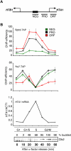
Figure 1.
Figure 1.
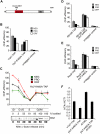
Yta7 and RNAPII binding to HTA1 chromatin is cell cycle-regulated. (A) Schematic representation of the PCR products (NEG, promoter [PRO], and ORF) used to cover the HTA1 locus in ChIP assays (Ng et al. 2002). (B) Rpb3 and Yta7 cross-link to HTA1 in a cell cycle-dependent manner. Rpb3-TAP and Yta7-TAP strains were arrested in G1 phase with 5 μM α-factor and released into fresh medium, and samples were taken at the indicated time points. IgG-sepharose ChIPs from the Rbp3-TAP or Yta7-TAP strains were analyzed for HTA1 (NEG, PRO, and ORF) by quantitative RT–PCR (qPCR), as indicated in the relevant panels. For each time point from the Yta7-TAP strain, cDNA was prepared and the ratio of HTA1 transcript to that of ACT1 was determined using qPCR (HTA1 mRNA panel). ChIP efficiency was calculated as described in the Materials and Methods. Cell cycle progression was monitored by assessing endogenous Clb2 cyclin levels (peak in G2/M phase) in the same samples by Western blotting with anti-Clb2 antibody and by quantification of budding (% budded). Similar results were seen for HTA1 transcript and Clb2 protein levels and budding indices in the Rpb3-TAP experiments (data not shown). Error bars in the experiments represent standard deviations from the mean for at least three replicate qPCR reactions.
Figure 2.
Figure 2.
Yta7 functionally interacts with RNAPII. (A) Yta7 interacts with RNAPII. Western blotting was used to probe an mChIP (Lambert et al. 2009) from Yta7-TAP or untagged cells with an antibody that recognizes RNAPII. (B) Affinity purification and identification of Yta7-TAP-associated proteins. A Coomassie-stained SDS–polyacrylamide gel is shown with affinity-purified proteins from Yta7-TAP and an untagged wild-type control; (Rb IgG) rabbit IgG. Copurifying proteins were identified by LC-MS/MS as described previously (Lambert et al. 2009), and the numbers of the unique peptides for each interacting protein are listed; (UC) untagged control. (C) RNAPII association with HTA1 chromatin is partly dependent on Yta7. ChIP analyses from logarithmically growing cells were performed as described in Figure 1B using the indicated strains and primer sets (PRO and ORF). Error bars in the experiments represent standard deviations from the mean for at least three replicate qPCR reactions.
Figure 3.
Figure 3.
The AAA-ATPase domain is required for the boundary function of Yta7. (A) Schematic representation of full-length Yta7. The AAA-ATPase domain (AAA-AD) and bromodomain (BD) are depicted, and the putative catalytic active lysine residue (amino acid 460) is indicated by an asterisk. (B) Localization of Yta7-K460A at the HTA1 locus. ChIP analyses (primer set PRO, NEG, and ORF) with samples from log-phase Yta7-TAP and Yta7-K460A-TAP and an untagged wild-type strain as a negative control were performed as described in Figure 1B. (C) Localization of Yta7-K460A to the HTA1 locus throughout the cell cycle. Yta7-K460A-TAP mutants were arrested in G1 phase with 5 μM α-factor and released into fresh medium, and ChIP analyses were performed as in Figure 1B using primer sets NEG, PRO, and ORF. Cell cycle progression was monitored by quantification of budding (entry into S phase) and by assessing endogenous Clb2 cyclin levels (peak in G2/M phase). (D) Localization of Rtt106 to the HTA1 promoter is dependent on the AAA-ATPase site. ChIP analyses with Rtt106-TAP in wild-type and yta7-K460A backgrounds from log-phase cultures were performed as described in Figure 1B (primer sets NEG, PRO, and ORF). An untagged wild-type strain was used as a control. (E) Localization of RSC to the HTA1 promoter is dependent on the AAA-ATPase site in Yta7. ChIP analyses with Rsc8-TAP in wild-type and yta7-K460A strains from log-phase cultures were performed as described in Figure 1B (primer sets NEG, PRO, and ORF). An untagged wild-type strain was used as a control. (F) Mutation of Lys 460 results in reduced HTA1 transcription, which is suppressed by elimination of Rtt106. cDNA was prepared from log-phase YTA7-TAP wild type, yta7-K460A-TAP and rtt106Δ single mutants, and a yta7-K460A-TAP rtt106Δ double mutant, and HTA1 transcript levels were assessed as described in Figure 1B. Error bars in the experiments represent standard deviations from the mean for at least three replicate qPCR reactions.
Figure 4.
Figure 4.
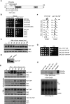
Yta7 is hyperphosphorylated by S-phase forms of Cdk1 and CK2. (A) Schematic representation of Cdk1 and CK2 phosphorylation sites in the N-terminal region of Yta7. (B) Growth defect caused by overexpression of YTA7 in the absence of Cdk1 G1–S-phase cyclins or CK2 subunits. Isogenic wild-type, cln1cln2, clb3clb4, clb5clb6, cln2clb5clb6, cka1, cka2, ckb1, or ckb2 deletion strains bearing either GAL-YTA7 (Hu et al. 2007) or empty vector were spotted in serial 10-fold dilutions on medium containing galactose and incubated for 2 d at 30°C. (C) Yta7 is phosphorylated in S phase. The phosphorylation of Yta7-TAP during a cell cycle was monitored by Western blotting using anti-TAP antibody. Cells were synchronized using 5 μM α-factor and released into fresh medium, and proteins extracts were prepared from samples taken at the indicated time points. Progression through the cell cycle was monitored by Western blotting for Clb2 (G2/M). Hxk1 was used as a gel loading control. (D) Mobility shift of Yta7 is due to phosphorylation. Yta7-TAP was isolated from wild-type cells in S phase and treated with λ-phosphatase (λ-PPase). (E) Phosphorylation of Yta7 is dependent on S-phase forms of Cdk1 and CK2 in vivo. (First panel) All experiments were performed under the same conditions and compared with wild type. (Second panel) Phosphoisoforms of Yta7-TAP were monitored during a cell cycle in a cln2Δclb5Δclb6Δ triple mutant exactly as described in C. (Third panel) Phosphorylation of Yta7-TAP after treatment with a CK2 inhibitor. Cells were synchronized as described in C and released into fresh medium supplemented with 100 mM CK2 inhibitor 4,5,6,7-tetrabromobenzotriazole (TBB) (Siepe and Jentsch 2009). Yta7-TAP, Clb2, and Hxt1 protein levels were assessed as described in C. (Fourth panel and F) Phenotypic assessment of a Yta7 phosphomutant. Phosphorylation of Yta7-13A-TAP was monitored during a cell cycle by Western blotting using anti-TAP antibody (Yta7-13A has all potential Cdk1 and CK2 sites [see A] converted to alanines). (Fourth panel) Samples were taken at the times indicated following α-factor block and release. (F) Corresponding FACS profiles indicate relative position in the cell cycle for the yta-13A-TAP strain and an isogenic wild-type control (YTA7-TAP). The arrow highlights the delayed mitosis in the yta7-13A mutant compared with wild type. (G) Growth defect caused by overexpression of yta7-13A. Wild-type, yta7-6A (lacking CK2 phospho-sites), yta7-7A (lacking Cdk1 phospho-sites) and yta7-13A (lacking both) strains in which the endogenous YTA7 promoter was replaced by the GAL1 promoter were spotted in serial 10-fold dilutions on glucose- or galactose-containing medium and incubated for 2 d at 30°C. (H) Phosphorylation of Yta7 by Cdk1 in vitro. (Top panel) Purification of Yta7-TAP, Yta7-6A-TAP, Yta7-7A-TAP, and Yta7-13A-TAP proteins from yeast was monitored by SDS-PAGE and silver staining. The purified proteins were incubated with Cln2–Cdk1 in kinase reactions along with [32P]-γ-ATP. (Bottom panel) Phosphorylation of proteins was analyzed by SDS-PAGE and autoradiography. The positions of migration of phosphorylated Yta7, Cln2, and Cdk1 are indicated. The asterisk marks the position of migration of a contaminant in the Cln2–Cdk1 preparation that is also phosphorylated in the reaction.
Figure 5.
Figure 5.
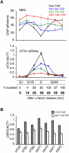
Dissociation of Yta7 from HTA1 chromatin ensures efficient transcription. (A) Phosphorylation of Yta7 regulates dissociation from HTA1 during S phase, which is essential for efficient HTA1 gene transcription. (Top panel) Yta7-TAP, Yta7-6A-TAP, Yta7-7A-TAP, and Yta7-13A-TAP strains were arrested with α-factor and released into fresh medium, and samples were taken at the time points indicated. ChIP analyses (primer set NEG) were performed as described in Figure 1B. (Bottom panel) S-phase induction of HTA1 transcription is markedly reduced in Yta7 phosphomutants. Samples of the same cultures used for the ChIP analyses were used for RNA isolation. cDNA was prepared from the samples, and HTA1 transcript levels were assessed as described in Figure 1B. Cell cycle progression was monitored by quantification of budding (entry into S phase). The budding indices for the Yta7-TAP strain are shown. S-phase entry was comparable for all strains (data not shown). (B) Analysis of transcript levels for other histone genes in a yta7 phosphomutant. YTA7-TAP and yta7-13A-TAP stains were arrested with α-factor and released into fresh medium, and samples were taken after 30 min (S phase). cDNA was prepared and analyzed as described in Figure 1B. Error bars in the experiments represent standard deviations from the mean for a least three replicate qPCR reactions.
Figure 6.
Figure 6.
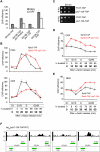
Phosphorylation of Yta7 is involved in transcript elongation by RNAPII. (A) Localization of Rtt106 in Yta7 mutants. Rtt106 localization to the HTA1 regulatory region (NEG) and ORF was assessed using ChIP in samples from asynchronous cultures of Rtt106-TAP strains harboring wild-type YTA7, yta7-K460A, yta7-13A, or both yta7-K460A and yta7-13A. ChIP analyses were performed as described in Figure 1B. (B) Analysis of RNAPII association with HTA1 during the cell cycle in a yta7-13A mutant. Rpb3-TAP localization to the promoter (primer set “PRO”) (top panel) and the ORF (primer set “ORF”) (bottom panel) of HTA1 was assayed in the indicated strains using qPCR as described (Fig. 1B). Cell cycle progression was monitored by quantification of budding (entry into S phase). Budding indices for the Rpb3-TAP strain are shown. S-phase entry was comparable for both strains (data not shown). (C) Sensitivity of yta7-13A to 6-azauracil. YTA7 and yta7-13A strains transformed with vector pRS-316 (for URA+ selection) were spotted in serial 10-fold dilutions on SD-URA plates with or without 6-azauracil (75 μg/mL) and incubated for 2 d at 30°C. (D) Increased localization of FACT to the HTA1 coding region in a yta7-13A strain during the cell cycle. Spt16-TAP and Spt16-TAP in a yta7-13A background were arrested in G1 phase with 5 μM α-factor and then released into fresh medium. ChIP samples were analyzed as described in Figure 1B (primer set ORF). Cell cycle progression was monitored by quantification of budding (entry into S phase). Budding indices for a Spt16-TAP strain are shown. S-phase entry was comparable for both strains (data not shown). (E) Increased localization of RSC to the HTA1 NEG region in a yta7-13A strain. Rsc8-TAP and Rsc8-TAP in yta7-13A were treated exactly as in D. ChIP samples were analyzed using primer set PRO. (F) Altered nucleosome occupancy in a yta7-13A mutant. Intergenic regions of all histone promoters are shown. A genome-wide nucleosome positioning assay was performed to identify regions of increased nucleosome occupancy in yta7-13A. Each track shows the change in nucleosome occupancy between mutant and wild type across the histone gene loci, expressed as the normalized log2 ratio of probe intensities.
Figure 7.
Figure 7.
Summary of Yta7 action and regulation. Yta7 acts as a boundary element during early G1, G2, and M phases. In S phase, Yta7 is phosphorylated by Cdk1 and CK2, which in turn is important for effective elongation of RNAPII along the HTA1 gene. See the text for details.

References

    1. Albuquerque CP, Smolka MB, Payne SH, Bafna V, Eng J, Zhou H 2008. A multidimensional chromatography technology for in-depth phosphoproteome analysis. Mol Cell Proteomics 7: 1389–1396 - PMC - PubMed
    1. Belotserkovskaya R, Oh S, Bondarenko VA, Orphanides G, Studitsky VM, Reinberg D 2003. FACT facilitates transcription-dependent nucleosome alteration. Science 301: 1090–1093 - PubMed
    1. Biswas D, Yu Y, Prall M, Formosa T, Stillman DJ 2005. The yeast FACT complex has a role in transcriptional initiation. Mol Cell Biol 25: 5812–5822 - PMC - PubMed
    1. Bloom J, Cross FR 2007. Multiple levels of cyclin specificity in cell-cycle control. Nat Rev Mol Cell Biol 8: 149–160 - PubMed
    1. Breitkreutz A, Choi H, Sharom JR, Boucher L, Neduva V, Larsen B, Lin ZY, Breitkreutz BJ, Stark C, Liu G, et al. 2010. A global protein kinase and phosphatase interaction network in yeast. Science 328: 1043–1046 - PMC - PubMed

Publication types

MeSH terms