. 2012 Apr;17(4):509-18.
doi: 10.1177/1087057111430253. Epub 2011 Dec 7.

A human islet cell culture system for high-throughput screening

Affiliations

A human islet cell culture system for high-throughput screening

Deepika Walpita et al. J Biomol Screen. 2012 Apr.

Abstract

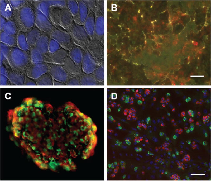
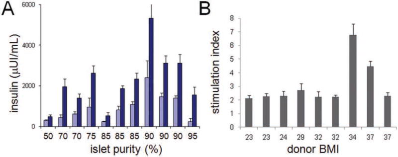
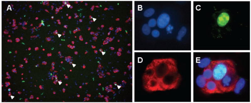
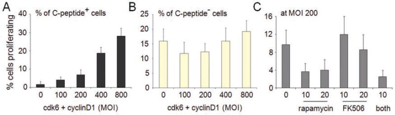
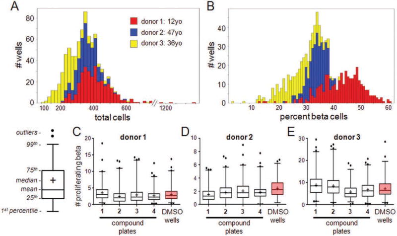
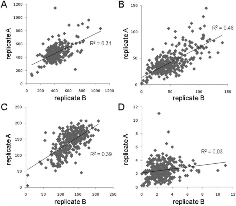
A small-molecule inducer of beta-cell proliferation in human islets represents a potential regeneration strategy for treating type 1 diabetes. However, the lack of suitable human beta cell lines makes such a discovery a challenge. Here, we adapted an islet cell culture system to high-throughput screening to identify such small molecules. We prepared microtiter plates containing extracellular matrix from a human bladder carcinoma cell line. Dissociated human islets were seeded onto these plates, cultured for up to 7 days, and assessed for proliferation by simultaneous Ki67 and C-peptide immunofluorescence. Importantly, this environment preserved beta-cell physiological function, as measured by glucose-stimulated insulin secretion. Adenoviral overexpression of cdk-6 and cyclin D(1), known inducers of human beta cell proliferation, was used as a positive control in our assay. This induction was inhibited by cotreatment with rapamycin, an immunosuppressant often used in islet transplantation. We then performed a pilot screen of 1280 compounds, observing some phenotypic effects on cells. This high-throughput human islet cell culture method can be used to assess various aspects of beta-cell biology on a relatively large number of compounds.


Conflict of interest statement

Declaration of Conflicting Interests

The authors declared no potential conflicts of interest with respect to the research, authorship, and/or publication of this article.

Figures

Figure 1
Figure 1
Preparation of surface for islet cell culture system. (A) Human bladder carcinoma HTB-9 cells grown to confluence on 96- and 384-well plates. Overlay of bright-field and fluorescent Hoechst nuclear dye. (B) After denuding the cells from the plate, the plate surface was stained for fibronectin (red) and laminin (green) levels. Scale bar = 10 µm. (C) Immunofluorescence on an intact human islet, revealing cellular architecture and distribution of cells. Red, insulin; green, glucagon. (D) Immunofluorescence on dissociated islet cells seeded in a 384-well plate. Red, C-peptide; green, glucagon; blue, Hoechst nuclear dye. Scale bar = 100 µm.
Figure 2
Figure 2
Cell culture system preserves glucose-stimulated insulin secretion (GSIS). (A) Islets from preparations with the indicated purity of isolation were dissociated, seeded into 96-well plates, and incubated for 1 h with either 1.67 mM (light blue bars) or 16.7 mM (dark blue bars) glucose to assess GSIS. (B) GSIS data are plotted relative to the body mass index (BMI) of the donor and, as the stimulation index, relative to 1.67 mM glucose. Data represent the mean ± standard deviation of four to eight independent wells.
Figure 3
Figure 3
Beta cell proliferation induced by cdk-6 and cyclin D1 in high-density culture. (A) Dissociated islet cells were cultured on extracellular matrix (ECM) and infected for 24 h with adenovirus vectors encoding cdk-6 and cyclin D1, incubated for 3 days in cell culture, and then fixed and assessed for C-peptide (a cleavage product of proinsulin) and Ki67 expression. Cells simultaneously positive for Ki67 and C-peptide are indicated by white arrowheads. To ensure that individual cells positive for Ki67 were expressing C-peptide, higher magnification images were acquired: (B) Hoechst nuclear dye, (C) Ki67, (D) C-peptide, and (E) image overlay.
Figure 4
Figure 4
Quantification of proliferation in response to cdk-6 and cyclin D1 . Dissociated islet cells infected for 24 h with increasing total multiplicity of infection (MOI) of cdk6 and cyclin D1 and incubated for 3 days were imaged and quantified for cell identity and proliferation status. (A) The percentage of proliferating C-peptide-expressing beta cells was quantified in each well of a 96-well plate. (B) The percentage of proliferating C-peptide-negative cells (including other islet endocrine cell types, endothelial cells, and fibroblasts) was quantified in each well of a 96-well plate. (C) Islet cells were infected with cdk-6 and cyclin D1 at an MOI of 200 for each gene in the absence or presence of indicated concentrations of the immunosuppressants rapamycin, FK506, or both. After a 3-day incubation, cells were fixed, stained, and quantified in the same manner as above. Data represent the mean ± standard deviations of 32 independent wells.
Figure 5
Figure 5
Pilot screen of 1280 compounds across three batches of islets. Islets from three organ donors were dissociated and cells seeded into extracellular matrix (ECM)–prepared 384-well plates. Cells were treated with individual members of a small-molecule collection, one compound per well, and incubated for 3 days. (A) Histogram of total cells per well across the three samples. (B) Histogram of the percentage of C-peptide-positive beta cells per well across the three samples. (C–E) Screening outcomes. The absolute number of proliferating beta cells was quantified in each well. Data are represented as box-whisker plots summarizing the wells of each compound plate (“1–4”) and all DMSO control wells across the plates (“DMSO wells,” boxed in pink). For the box-whisker plots, the mean value of the data set is represented by the central line of the box and the median by “+.” The box size represents the 25th and 75th percentiles of the data and the whiskers the 1st and 99th percentiles. Further outliers are represented by dots. Graphical schematic is shown. Box-whisker plots of screening outcomes for (C) donor 1, (D) donor 2, and (E) donor 3.
Figure 6
Figure 6
Analysis of screening performance reproducibility. Islet cells were dissociated and treated for 3 days in duplicate with 1280 compounds. The two replicates are plotted against each other examining (A) total cells, (B) proliferating C-peptide-negative cells, (C) quiescent C-peptide-positive beta cells, and (D) proliferating beta cells. The correlations of each data set to a linear fit are shown.

References

    1. Shapiro AM, Lakey JR, Ryan EA, Korbutt GS, Toth E, Warnock GL, Kneteman NM, Rajotte RV. Islet Transplantation in Seven Patients with Type 1 Diabetes Mellitus Using a Glucocorticoid-Free Immunosuppressive Regimen. N. Engl. J. Med. 2000;343:230–238. - PubMed
    1. Shapiro AM, Ricordi C, Hering BJ, Auchincloss H, Lindblad R, Robertson RP, Secchi A, Brendel MD, Berney T, Brennan DC, et al. International Trial of the Edmonton Protocol for Islet Transplantation. N. Engl. J. Med. 2006;355:1318–1330. - PubMed
    1. Meier JJ, Bhushan A, Butler AE, Rizza RA, Butler PC. Sustained Beta Cell Apoptosis in Patients with Long-Standing Type 1 Diabetes: Indirect Evidence for Islet Regeneration? Diabetologia. 2005;48:2221–2228. - PubMed
    1. Meier JJ, Lin JC, Butler AE, Galasso R, Martinez DS, Butler PC. Direct Evidence of Attempted Beta Cell Regeneration in an 89-Year-Old Patient with Recent-Onset Type 1 Diabetes. Diabetologia. 2006;49:1838–1844. - PubMed
    1. Keenan HA, Sun JK, Levine J, Doria A, Aiello LP, Eisenbarth G, Bonner-Weir S, King GL. Residual Insulin Production and Pancreatic Beta-Cell Turnover after 50 Years of Diabetes: Joslin Medalist Study. Diabetes. 2010;59:2846–2853. - PMC - PubMed

Publication types

MeSH terms

LinkOut - more resources