

Mice with mutations in Fas and Fas ligand demonstrate increased herpetic stromal keratitis following corneal infection with HSV-1
- PMID: 22156346
- PMCID: PMC3253206
- DOI: 10.4049/jimmunol.1102251
Mice with mutations in Fas and Fas ligand demonstrate increased herpetic stromal keratitis following corneal infection with HSV-1
Abstract
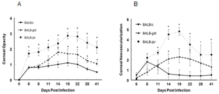
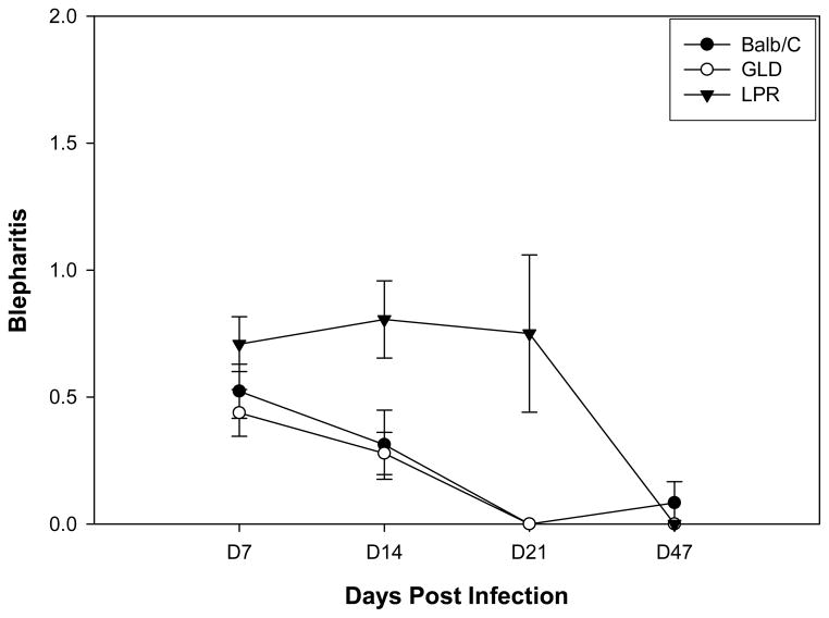
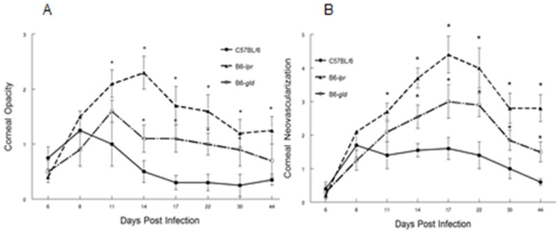
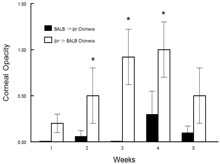
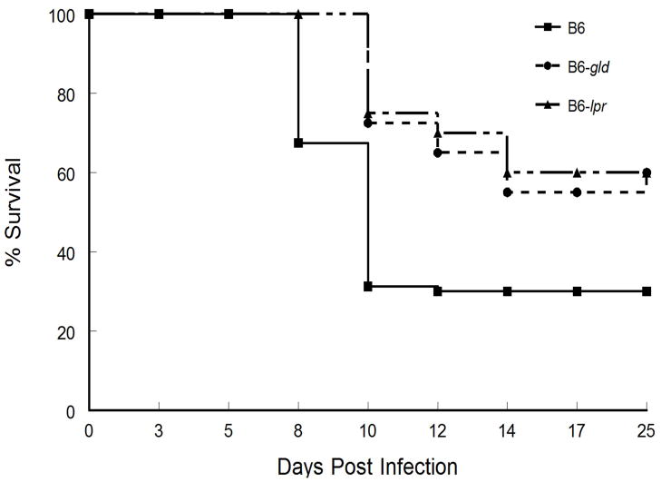
HSV-1 infection of the cornea leads to a potentially blinding immunoinflammatory lesion of the cornea, termed herpetic stromal keratitis. It has also been shown that one of the factors limiting inflammation of the cornea is the presence of Fas ligand (FasL) on corneal epithelium and endothelium. In this study, the role played by FasL expression in the cornea following acute infection with HSV-1 was determined. Both BALB/c and C57BL/6 (B6) mice with HSV-1 infection were compared with their lpr and gld counterparts. Results indicated that mice bearing mutations in the Fas Ag (lpr) displayed the most severe disease, whereas the FasL-defective gld mouse displayed an intermediate phenotype. It was further demonstrated that increased disease was due to lack of Fas expression on bone marrow-derived cells. Of interest, although virus persisted slightly longer in the corneas of mice bearing lpr and gld mutations, the persistence of infectious virus in the trigeminal ganglia was the same for all strains infected. Further, B6 mice bearing lpr and gld mutations were also more resistant to virus-induced mortality than were wild-type B6 mice. Thus, neither disease nor mortality correlated with viral replication in these mice. Collectively, the findings indicate that the presence of FasL on the cornea restricts the entry of Fas(+) bone marrow-derived inflammatory cells and thus reduces the severity of HSK.
Figures
Similar articles
-
Recurrent herpetic stromal keratitis in mice: a model for studying human HSK.Clin Dev Immunol. 2012;2012:728480. doi: 10.1155/2012/728480. Epub 2012 Apr 23. Clin Dev Immunol. 2012. PMID: 22593769 Free PMC article. Review.
-
Impaired Fas-Fas Ligand Interactions Result in Greater Recurrent Herpetic Stromal Keratitis in Mice.J Immunol Res. 2015;2015:435140. doi: 10.1155/2015/435140. Epub 2015 Oct 4. J Immunol Res. 2015. PMID: 26504854 Free PMC article.
-
Effect of Fas and Fas ligand deficiency in resistance of C57BL/6 mice to HSV-1 keratitis and chorioretinitis.Invest Ophthalmol Vis Sci. 2001 Oct;42(11):2505-9. Invest Ophthalmol Vis Sci. 2001. PMID: 11581190
-
Fas and Fas ligand expressed on cells of the immune system, not on the target tissue, control induction of experimental autoimmune uveitis.J Immunol. 2000 Nov 15;165(10):5480-6. doi: 10.4049/jimmunol.165.10.5480. J Immunol. 2000. PMID: 11067900
-
Role of Herpes Simplex Virus Type 1 (HSV-1) Glycoprotein K (gK) Pathogenic CD8+ T Cells in Exacerbation of Eye Disease.Front Immunol. 2018 Dec 7;9:2895. doi: 10.3389/fimmu.2018.02895. eCollection 2018. Front Immunol. 2018. PMID: 30581441 Free PMC article. Review.
Cited by
-
The immunoregulatory role of corneal epithelium-derived thrombospondin-1 in dry eye disease.Ocul Surf. 2018 Oct;16(4):470-477. doi: 10.1016/j.jtos.2018.07.005. Epub 2018 Jul 25. Ocul Surf. 2018. PMID: 30055331 Free PMC article.
-
SLURP-1 modulates corneal homeostasis by serving as a soluble scavenger of urokinase-type plasminogen activator.Invest Ophthalmol Vis Sci. 2014 Aug 28;55(10):6251-61. doi: 10.1167/iovs.14-15107. Invest Ophthalmol Vis Sci. 2014. PMID: 25168896 Free PMC article.
-
Inhibition of HUVEC tube formation via suppression of NFκB suggests an anti-angiogenic role for SLURP1 in the transparent cornea.Exp Eye Res. 2017 Nov;164:118-128. doi: 10.1016/j.exer.2017.08.007. Epub 2017 Aug 10. Exp Eye Res. 2017. PMID: 28803936 Free PMC article.
-
When Clarity Is Crucial: Regulating Ocular Surface Immunity.Trends Immunol. 2018 Apr;39(4):288-301. doi: 10.1016/j.it.2017.11.007. Epub 2017 Dec 14. Trends Immunol. 2018. PMID: 29248310 Free PMC article. Review.
-
Recurrent herpetic stromal keratitis in mice: a model for studying human HSK.Clin Dev Immunol. 2012;2012:728480. doi: 10.1155/2012/728480. Epub 2012 Apr 23. Clin Dev Immunol. 2012. PMID: 22593769 Free PMC article. Review.
References
-
- Pepose JS, Leib DA, Stuart PM, Easty EL. Herpes simplex virus diseases: anterior segment of the eye. In: Pepose JS, Holland GAN, Wilhelmus KR, editors. Ocular Infection and Immunity. Mosby; St. Louis, MO: 1996. pp. 905–932.
-
- Thomas J, Gangappa S, Kanangat S, Rouse BT. On the essential involvement of neutrophils in the immunopathologic disease: herpetic stromal keratitis. J Immunol. 1997;158:1383–1391. - PubMed
-
- Maertzdorf J, Verjans GM, Remeijer L, van der Kooi A, Osterhaus AD. Restricted T cell receptor beta-chain variable region protein use by cornea-derived CD4+ and CD8+ herpes simplex virus-specific T cells in patients with herpetic stromal keratitis. J Infect Dis. 2003;187:550–558. - PubMed
-
- Denniston AK, Kottoor SH, Khan I, Oswal K, Williams GP, Abbott J, Wallace GR, Salmon M, Rauz S, Murray PI, Curnow SJ. Endogenous cortisol and TGF-beta in human aqueous humor contribute to ocular immune privilege by regulating dendritic cell function. J Immunol 2011. 2011;186:305–311. - PubMed
Publication types
MeSH terms
Substances
Grants and funding
LinkOut - more resources
Full Text Sources
Molecular Biology Databases
Research Materials
Miscellaneous
