. 2012 Apr;40(7):3159-71.
doi: 10.1093/nar/gkr1228. Epub 2011 Dec 9.

New function for the RNA helicase p68/DDX5 as a modifier of MBNL1 activity on expanded CUG repeats

Affiliations

New function for the RNA helicase p68/DDX5 as a modifier of MBNL1 activity on expanded CUG repeats

François-Xavier Laurent et al. Nucleic Acids Res. 2012 Apr.

Abstract

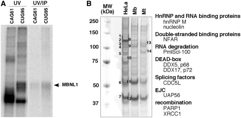
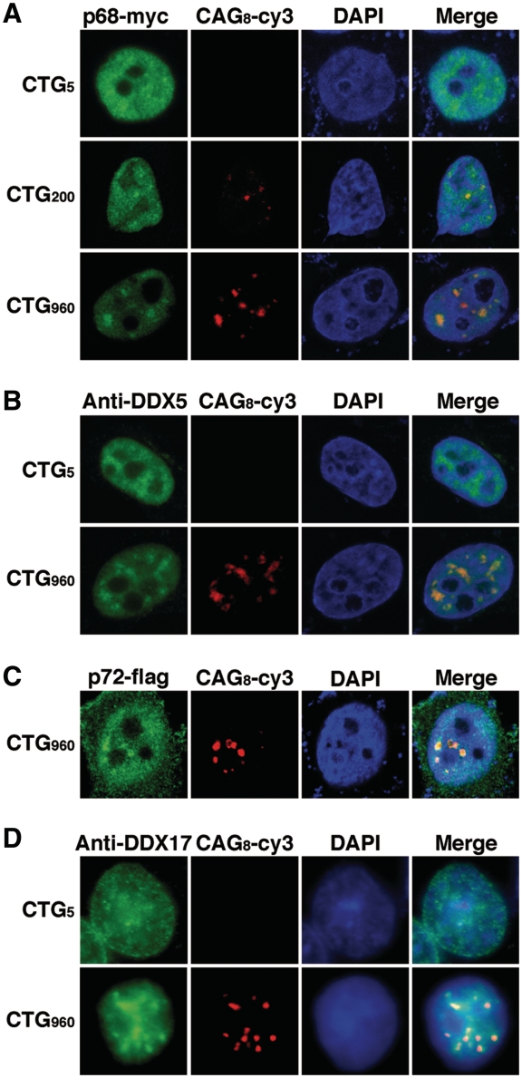
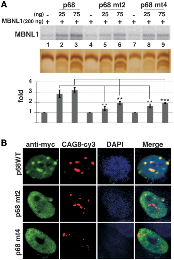
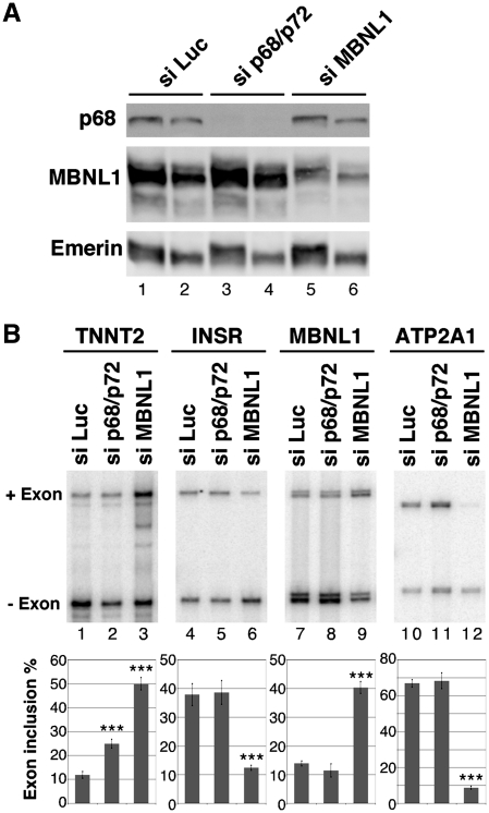
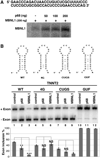
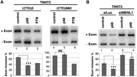
Myotonic Dystrophy type I (DM1) is caused by an abnormal expansion of CTG triplets in the 3' UTR of the dystrophia myotonica protein kinase (DMPK) gene, leading to the aggregation of the mutant transcript in nuclear RNA foci. The expanded mutant transcript promotes the sequestration of the MBNL1 splicing factor, resulting in the misregulation of a subset of alternative splicing events. In this study, we identify the DEAD-box RNA helicase p68 (DDX5) in complexes assembled onto in vitro-transcribed CUG repeats. We showed that p68 colocalized with RNA foci in cells expressing the 3'UTR of the DMPK gene containing expanded CTG repeats. We found that p68 increased MBNL1 binding onto pathological repeats and the stem-loop structure regulatory element within the cardiac Troponin T (TNNT2) pre-mRNA, splicing of which is misregulated in DM1. Mutations in the helicase core of p68 prevented both the stimulatory effect of the protein on MBNL1 binding and the colocalization of p68 with CUG repeats, suggesting that remodeling of RNA secondary structure by p68 facilitates MBNL1 binding. We also found that the competence of p68 for regulating TNNT2 exon 5 inclusion depended on the integrity of MBNL1 binding sites. We propose that p68 acts as a modifier of MBNL1 activity on splicing targets and pathogenic RNA.


Figures

Figure 1.
Figure 1.
Affinity capture of protein complexes from HeLa and myogenic nuclear extracts by an in vitro transcribed RNA containing 95 CUG repeats. (A) MBNL1 is cross-linked to 32P-labeled CUG95 repeats. In vitro transcribed (CUG)95 or (CAG)61 RNAs labelled with [α-32P]CTP were incubated with 40% HeLa nuclear extracts. After UV cross-linking an aliquot was precipitated with anti-MBNL1 antibodies. The cross-linked proteins were separated by SDS-PAGE. (B) Proteins from HeLa nuclear extracts, myoblast (Mb) and myotube (Mt) nuclear extracts from C2C12 were separated onto a 10% polyacrylamide denaturing gel and detected by Coomassie Brilliant Blue staining. Numbers 1 through 14 refer to bands that have been cut out from the gel and identified by mass-spectrometry. A subset of proteins identified by mass spectrometry is indicated.
Figure 2.
Figure 2.
p68 and p72 colocalized with CUG RNA foci in HeLa cells. (A) Cells were cotransfected with a plasmid encoding p68-myc and plasmids expressing either 5, 200 or 960 CTG repeats. Expression of p68-myc was detected by immunofluorescence using monoclonal anti-Myc antibodies, followed by rabbit anti-mouse A488 antibodies. Foci were detected by RNA-FISH using Cy3-labeled (CAG)8 probes. Nuclei were stained with DAPI. (B) Endogenous p68 colocalizes with CUG repeats. p68/DDX5 was revealed with a DDX5 monoclonal antibody. (C) Same experiment as in (A) except that cells were cotransfected with a Flag-p72 expression vector. Expression of Flag-p72 was detected by immunofluorescence using anti-Flag antibodies. (D) Endogenous p72 colocalizes with CUG repeats. P72/DDX17 was revealed with DDX17 polyclonal antibodies.
Figure 3.
Figure 3.
p68 increases the binding of MBNL1 to 95 CUG repeats. (A) Plasmid (CUG)95 was in vitro transcribed in the presence of [α-32P]UTP. Labeled (CUG)95 RNA was incubated with a constant amount of recombinant MBNL1 protein (200 ng) and increasing amounts of recombinant p68ΔCt2 under splicing conditions with ATP. Proteins cross-linked to labeled RNA were separated on a 10% SDS–PAGE. Bottom image shows a silver stain of a gel run in parallel. Note that p68 ΔCt2 migrates as two bands that are recognized by anti-p68 antibodies (data not shown). (B) The cross-linked proteins shown in (A) were immunoprecipitated with anti-MBNL1 antibodies and separated by SDS-PAGE. (C) (CUG)95, (CAG)61 and (CCUG)62 RNAs were labeled with [α-32P]CTP and used for UV-cross-linking experiments. (D) The increase of cross-linked MBNL1 to CUG repeats is specific to p68. Labeled (CUG)95 was incubated with 200 ng of recombinant MBNL1 protein and 25 or 75 ng of recombinant p68ΔCt2, UAP56 or eIF4A3 proteins. Quantifications result from three independent experiments, with error bars indicating standard deviation. *P < 0.05, **P < 0.01, ***P < 0.001.
Figure 4.
Figure 4.
(A) Mutations in the helicase core domain of p68 modify its ability to increase MBNL1 binding. UV-cross-linking experiments with labeled (CUG)95 were performed with 200 ng of recombinant MBNL1 and 25 or 75 ng of wild-type recombinant p68ΔCt2 or p68ΔCt2 mutated in domain II (p68 mt2, mutation D248N), or in domain IV (p68 mt4, mutation F346A). Quantifications result from three independent experiments, with error bars indicating standard deviation. *P < 0.05, **P < 0.01, ***P < 0.001. (B) Mutations in the helicase core domain of p68 strongly affect colocalization of the protein with CUG repeats. RNA/FISH experiments were performed as described in Figure 2 with wild-type p68-myc expressing vector or p68-myc expressing vectors mutated in domain II (DEAD) or IV.
Figure 5.
Figure 5.
P68 and p72 facilitate RNA foci formation. Human muscle cells expressing 960-interrupted CTG repeats under the control of a Tet-on inducible promoter was used to attend the early steps of RNA foci formation. The number of nuclei containing foci was determined at different times after doxycycline addition (1, 3, 6 and 24 h) in non-treated or treated cells by siRNA to inhibit p68/p72. SiRNA duplexes were transfected 48 h before CUG repeats induction by doxycycline. Quantification results from three independent experiments. Approximately 300 cells were counted for each condition, with error bars indicating standard deviation *P < 0.05, **P < 0.01, ***P < 0.001.
Figure 6.
Figure 6.
p68 regulates splicing of TNNT2 alternative exon 5. (A) Western blot analysis of human myoblasts treated with a control luciferase siRNA, (siLuc), sip68/p72 or siMBNL1. About 15 µg (lanes 1, 3 and 5) or 7.5 µg (lanes 2, 4 and 6) of proteins were loaded per lane. The blot was probed with antibodies against p68, MBNL1 and emerin as a control. (B) RT–PCR analysis of endogenous transcripts in human myoblast cells transfected with a control siRNA (siLuc), siRNA against p68/p72 or siRNA against MBNL1. The histogram shows the quantification of exon inclusion. Results are derived from six independent experiments for TNNT2 and four independent experiments for INSR, MBNL1 and ATP2A1 with error bars indicating standard deviation. *P < 0.05, **P < 0.01, ***P < 0.001.
Figure 7.
Figure 7.
RNA stem–loop mutations that abolish MBNL1 binding impair the regulation by p68. (A) p68 stimulates binding of MBNL1 onto the stem–loop regulatory element of TNNT2. Labeled stem–loop regulatory RNA was incubated with a constant amount of recombinant MBNL1 protein (200 ng) and increasing amounts of recombinant p68ΔCt2. Proteins cross-linked to labeled RNA were separated on a 10% SDS–PAGE. The TNNT2 RNA used for cross-linking experiments is indicated. (B) RT–PCR analysis of splicing of TNNT2 alternative exon 5 in wild-type and mutant minigenes. Schematic representation of wild-type and mutant stem–loop structure within TNNT2 intron 4 [adapted from ref. (31)] is shown. Human myoblast cells were cotransfected with wild-type or mutant TNNT2 minigenes and expression vector coding for p68 (lanes 2, 5, 8 and 11) or MBNL1 (lanes 3, 6, 9 and 12). The histogram shows the quantification of exon 5 inclusion. Results are derived from at least three experiments, except for lanes 4, 6, 7, 9, 10 and 12, which are derived from two experiments. *P < 0.05, **P < 0.01, ***P < 0.001. NS: non-specific.
Figure 8.
Figure 8.
Sequestration of MBNL1 by CUG repeats or MBNL1 knockdown by siRNA makes splicing of TNNT2 exon 5 non-responsive to p68. (A) RT–PCR analysis of TNNT2 minigene in human myoblast cells cotransfected with the 3′ UTR DMPK minigene containing 5 (lanes 1–3) or 960 (lanes 4 to 6) CTG repeats and a p68 expression vector (lanes 2 and 5) or as a control a PTB expression vector (lanes 3 and 6). The histogram shows the quantification of exon 5 inclusion. Results are from at least two experiments except lanes 3 and 6, for which a single experiment was performed. (B) RT–PCR analysis of TNNT2 minigene in human myoblast cells transfected with a control siRNA (lanes 1 and 2) or siRNA against MBNL1 (lanes 3 and 4) and a p68 expression vector (lanes 2 and 4). The histogram shows the quantification of exon 5 inclusion. Results are from four experiments with error bars indicating standard deviation. *P < 0.05, **P < 0.01, ***P < 0.001.

Similar articles

Cited by

References

    1. Harper PS. Myotonic Dystrophy. London: W. B. Saunders; 2001.
    1. Brook JD, McCurrach ME, Harley HG, Buckler AJ, Church D, Aburatani H, Hunter K, Stanton VP, Thirion JP, Hudson T, et al. Molecular basis of myotonic dystrophy: expansion of a trinucleotide (CTG) repeat at the 3′ end of a transcript encoding a protein kinase family member. Cell. 1992;68:799–808. - PubMed
    1. Mahadevan M, Tsilfidis C, Sabourin L, Shutler G, Amemiya C, Jansen G, Neville C, Narang M, Barcelo J, O'Hoy K, et al. Myotonic dystrophy mutation: an unstable CTG repeat in the 3' untranslated region of the gene. Science. 1992;255:1253–1255. - PubMed
    1. Fu YH, Pizzuti A, Fenwick RG, Jr, King J, Rajnarayan S, Dunne PW, Dubel J, Nasser GA, Ashizawa T, de Jong P, et al. An unstable triplet repeat in a gene related to myotonic muscular dystrophy. Science. 1992;255:1256–1258. - PubMed
    1. Taneja KL, McCurrach M, Schalling M, Housman D, Singer RH. Foci of trinucleotide repeat transcripts in nuclei of myotonic dystrophy cells and tissues. J. Cell. Biol. 1995;128:995–1002. - PMC - PubMed

Publication types